
selection guide
Cleaning an amplicon before sequencing
Choose a column, bead or gel cleanup so leftover primers and dNTPs do not wreck the sequencing read you are about to request.
- Author
- EVRINTH Editorial Team
- Published
- 8 October 2026
- Updated
- 8 October 2026
- Reading time
- 7 min
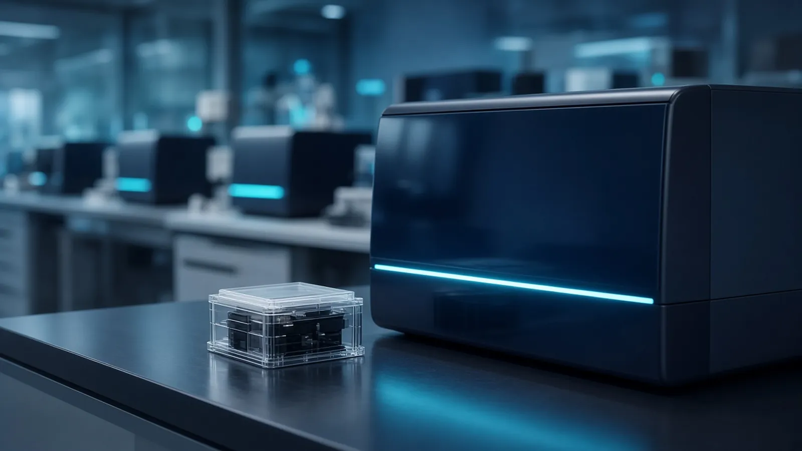
Leftover primers and dNTPs in a finished PCR will poison a Sanger reaction. The selection this guide supports is among three cleanup classes: a silica column, magnetic beads, or cutting one band out of a gel. Choose from what the gel showed and from which instrument will read the DNA. The reaction that made the amplicon is described in how polymerase chain reaction works. What a sequencing provider needs before the run starts is in what to check before a sequencing run. If the destination is a library on a flow cell, the path is next-generation sequencing from library to reads, and the cleanup follows that library kit.
The photograph is a benchtop sequencing instrument with a teal status light and a flow cell cartridge. There is no person in that frame, and the machine is not a Sanger capillary array. Keep that distinction when you choose a cleanup. Columns, beads and gel slices are reagent classes you can specify from the molecular biology catalogue and the quote request.
What you are trying to remove
A completed PCR still contains the primers you added, the dNTPs the polymerase did not use, salts, and often the enzyme. Sanger sequencing primes again from a single oligo and incorporates dye-labelled terminators in competition with ordinary dNTPs. Extra primers give extra start sites. Extra dNTPs dilute the terminators. The chromatogram comes back noisy, short, or double. A bright gel band does not mean those small molecules are gone. They are too small to see as the band.
Cleanup also concentrates the amplicon into a buffer the sequencing reaction tolerates. Water or a low-salt elution is the usual target. Follow the cleanup product you opened for elution volume. Do not invent a microlitre table here.
Before you clean, look at the gel beside a marker, with the no-template control on the same image. One band at the expected size, and a clean blank, is the condition for a column or a bead. Two bands, or a blank that matches the sample, means you are not ready to sequence. A dirty blank is contamination. Sequencing it produces a confident read of the wrong molecule. Fix the PCR before you clean it. How to read that gel is the method in agarose gel electrophoresis for DNA.
Three classes, and the criterion for each
A silica column binds double-stranded DNA in a chaotropic salt, washes away small molecules, and elutes the DNA in low salt. Primers and free dNTPs leave in the wash if they are below the column's practical size retention. Very short amplicons can leave with them. The column insert states the length it still binds. Follow that insert. If your product is near the cutoff, a bead method with a ratio you have checked is often the safer class. Columns are a good default for a single band of ordinary length when you want a simple spin and a clean elution.
Magnetic beads of the solid-phase reversible kind bind DNA in a polyethylene glycol and salt mixture. A magnet replaces the spin. The ratio of bead suspension to sample changes the size that is kept. Higher ratios keep shorter fragments. That is useful when the amplicon is short, and it is a hazard when you needed to exclude a slightly shorter side product. The ratio is part of the selection, and it comes from the bead protocol, not from a guess. Beads also suit plate-scale cleanup. They still will not separate two bands that are both long enough to bind.
Gel extraction is the class you choose when the gel shows more than one product and you need a single sequence. You separate on agarose, cut the band you want, dissolve the slice, and bind the DNA to a column or to beads. You pay in yield, in time, and in damage if the band was visualised with ultraviolet light. Ultraviolet nicks DNA. Those nicks stall a sequencing polymerase and can appear as noise or early stops. Use a blue-light box when the stain is compatible, keep the exposure short if ultraviolet is the only option, and do not look at a bare ultraviolet surface. An orange shield is there to be used. The stain's own safety data still applies.
Enzymatic cleanup, a mix of exonuclease and phosphatase activities that chew primers and degrade unused dNTPs, is a fourth class some laboratories use when there is truly one product. It does not remove a second band. If you use it, follow that mix, and heat-kill it as the card requires so it does not chew the sequencing primer later. This guide treats column, bead and gel as the three selections to make first, because those are the ones that also change salt and buffer.
| What the gel and the instrument show | Cleanup class | What can still go wrong |
|---|---|---|
| One band, Sanger of that amplicon | Column or beads | Short products lost on a column with a high cutoff |
| One short amplicon near a column's limit | Beads, ratio checked for that length | A ratio that also keeps primer-dimer |
| Two or more bands, one of them wanted | Gel slice, then column or beads | Ultraviolet nicking if you cut on a UV box |
| Library for the flow-cell instrument in the photograph | The library kit's own bead or column step | Adapter-dimers that a Sanger column protocol was not asked to remove |
| No-template lane matches the sample | Do not clean for sequence yet | You would sequence the contaminant |
After the cleanup, before you ask for a read
Quantify what you eluted. Absorbance at 260 nanometres estimates nucleic acid and also sees free nucleotides you hoped you had removed. A dye selective for double-stranded DNA ignores those leftovers better and needs a standard curve. A260/A280 near 1.8 is a cleanliness hint for DNA, not proof of identity or of intact ends. If absorbance is high and a dye reading is low, the cleanup may have failed to remove small material, or the DNA may be single-stranded or damaged. The Addgene quantification note is a public method for that comparison. Follow your dye's standard curve.
Match the sequencing primer to one end of the amplicon. One Sanger read does not cover a long product. Say how far you need to see. Mixed peaks after a clean single band usually mean the cleanup was fine and the template was mixed, or the primer binds twice. Do not repeat the same dirty mixture and hope.
For the instrument in the photograph, stop and switch methods. A flow cell wants a library: fragmented or amplicon-tagged DNA, adapters, a size profile, and a cleanup aimed at adapter-dimer. Sending a Sanger-cleaned PCR tube to that instrument because both are "sequencing" wastes the flow cell. The library article is the decision path. The check-before-a-run article is the list of identity, amount and indexes to confirm.
Safety
Chaotropic salts used to bind DNA to silica are chemical hazards. Follow the cleanup product's safety data and your institutional waste rules. Ultraviolet light damages eyes, skin and DNA. Blue light is kinder to the fragment and still not a reason to skip eye protection the box requires. Research sequencing is not a diagnostic result. A clean chromatogram of a teaching amplicon does not become a clinical genotype.
What to send with a sequencing question
Send the amplicon length, a description of the gel (one band or a slice), the cleanup class you used, how you quantified and the approximate mass, the sequencing primer and which end it binds, and a reference accession if you have one in a public nucleotide record. Say whether the read is Sanger of a PCR product or a library for a flow-cell instrument. Say that a band was not treated as identity. The nucleic acid analysis pathway connects extraction, PCR and the read. Ask whether a quotation is possible for the cleanup reagents or for the sequencing question. The multiplex PCR enquiry reference is relevant only if several products were amplified on purpose and now need to be told apart. Use it as an enquiry reference.
Questions from the bench
Why do leftover primers and dNTPs spoil a Sanger reaction?
The sequencing reaction still uses a primer and a balance of ordinary nucleotides against dye-labelled terminators. Leftover PCR primers anneal and extend, so you get extra peaks or a noisy start. Leftover dNTPs change that terminator balance, which shortens or unbalances the read. Removing them is the point of the cleanup, not a polish step you can skip because the gel looked bright.
When is a column or a bead cleanup the wrong class?
When more than one product is visible and you need only one of them. A column or a bead step that keeps all double-stranded DNA above a size cutoff will keep the side band too, and Sanger will read the mixture as overlapping peaks. Gel extraction, or a redesign that yields one band, is the selection in that case.
What does ultraviolet light do if I cut the band out?
Ultraviolet light nicks DNA and damages eyes and skin. Nicks and lesions stop a sequencing polymerase or show up as odd peaks and weak signal. Minimise exposure, prefer a blue-light transilluminator when the stain allows, and do not look at a bare ultraviolet box. The orange shield on a gel imager exists for that reason.
The photograph shows a benchtop sequencer and a flow cell. Is this cleanup the same as a Sanger cleanup?
The instrument in the picture is a benchtop sequencer with a flow cell, which takes a library, not a raw PCR tube. Library cleanup follows that library kit and is aimed at adapter-dimers and size. Sanger cleanup of a single amplicon is the column, bead or gel choice above. Tell the sequencing enquiry which instrument class you mean so the cleanup matches it.
References
Manufacturer names identify published method classes. Trademarks remain with their owners. Catalogue records on this site are independent references for enquiry. They are not a statement of inventory, distribution rights or a supply commitment. This page is educational. It is not medical advice, a diagnostic protocol or a biosafety approval.
Catalogue
Related products and categories
These links follow the subject of the article into published manufacturer references. A listing is a reference for an enquiry, not a statement of stock or distribution rights.
Continue in this cluster
Related reading
 How polymerase chain reaction worksHow PCR copies a chosen DNA segment: the temperature cycle, reagents, controls and what a band on a gel does and does not prove.A glossary of PCR termsPlan a PCR by defining amplicon, Tm, Ta, hot-start, plateau, master mix, NTC, inhibition, fidelity and multiplex before you mix.Choosing an annealing temperatureWhy annealing temperature is not the Tm printed on a tube, and how salt, magnesium, additives, and a short gradient locate the real window.