
guide
Affinity and ion-exchange protein purification
How affinity capture and ion exchange pull a protein from a lysate, and why each collected fraction still needs an identity check.
- Author
- EVRINTH Editorial Team
- Published
- 8 October 2026
- Updated
- 8 October 2026
- Reading time
- 9 min
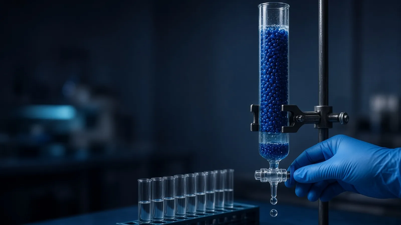
Affinity and ion-exchange protein purification are two preparative chromatography modes that answer different questions. Affinity asks whether the protein carries a feature a ligand recognises. Ion exchange asks what net charge that protein shows in a stated buffer. Used in series, they are how many laboratories take a recombinant protein from a clarified lysate to a fraction clean enough to assay. The decision this page supports is when to trust a binding step, when to add a charge step, and when a fraction is still only a fraction. The broader menu of modes, including reversed phase and size exclusion, is in chromatography methods in life-science labs.
Columns and instruments are compared from the scientific instruments catalogue. A campaign question can be framed against the custom protein expression and purification reference. That page does not mean EVRINTH is performing the purification. A specification goes to the quote request.
When this pair is the right pair
Use affinity first when the construct has a real handle: a histidine tag, a glutathione S-transferase domain, a maltose-binding domain, a biotinylated site, or an epitope an immobilised antibody binds. The step throws away most of the host proteome. Use ion exchange second when the affinity eluate still contains proteins that shared the handle, or when you must separate a full-length chain from a fragment that also stuck. Use ion exchange alone when there is no tag and you know the charge well enough to bind the target and wash the neighbours off. Use neither as the first step on a lysate so crude that lipid and debris will pack the frit. Clarify first.
Sequence is the planning input. UniProt records mass, domains and, where someone has measured or predicted it, isoelectric information you can use as a starting estimate. The estimate is not the behaviour in your buffer, with your tag, at your ionic strength. HUPO is a reminder that identifying the protein in the fraction is a proteomics claim with its own standards, not a peak label.
Affinity is a chosen binding event
Immobilised-metal affinity chromatography is the common histidine-tag method. A resin presents nickel, cobalt or another transition metal. Histidine side chains coordinate that metal. Low imidazole in the load and wash competes weakly and knocks off poor binders. Higher imidazole elutes the tagged protein. Cobalt resins are often more selective and less capacious than nickel resins. That is a trade, not a moral ranking. Chelators such as EDTA strip the metal and kill the step. Many protease-inhibitor tablets contain them. Read the lysis buffer.
Other affinity classes are the same logic with a different pair. Glutathione resin binds glutathione S-transferase fusions. Amylose binds maltose-binding protein. Streptavidin binds biotin with a strength that can make elution the hard part. Protein A or protein G binds the constant region of many antibodies and is a purification of the antibody, not of the antigen, unless you have built a different experiment. Elution is a competitor, a pH change, or a cleavage that cuts the tag and lets the protein leave while the partner stays. Cleavage is a second purification problem. The protease and the cut tag have to be removed, often by a second affinity pass that now binds what you no longer want.
Host proteins bind these resins too. E. coli proteins with surface histidines are the classic nickel-column contaminants. A tall 280-nanometre peak can be your protein, a contaminant, or both. The gel of the load, the flow-through, the wash and each elution fraction is the minimum picture. Activity, if you have an assay, is better. A peak that is active and dirty is still a useful intermediate. A peak that is clean and inactive may be a folded failure or the wrong protein.
Tags change the molecule. A histidine stretch, a fusion partner and a linker are part of the mass and sometimes part of the function until you remove them. Plan the tag with the assay, not after the column disappoints you.
Ion exchange is charge in a stated buffer
Anion exchangers such as quaternary ammonium or diethylaminoethyl resins are positive and bind negative proteins. Cation exchangers such as sulfopropyl or carboxymethyl resins are negative and bind positive proteins. Quaternary and sulfopropyl types stay charged over a wide pH range and are called strong. Diethylaminoethyl and carboxymethyl types change charge near their own pKa and are called weak. The adjective is about the ligand, not about how fiercely your protein sticks.
The isoelectric point is a guide to the starting pH. A buffer well above that point pushes many proteins negative and toward an anion exchanger. A buffer well below it does the opposite. Stay a pH unit away from the point if you want a clear net charge, and remember that the protein can precipitate at its isoelectric point. Also remember patches: a protein can stick to a cation exchanger above its isoelectric point because a positive face remains. A two-condition trial teaches more than a dogma.
Salt elutes by competition. A gradient from low salt to high salt releases weak binders first and strong binders later. A step elution is faster and usually less resolving. pH elution changes the protein's charge until it lets go. It is powerful and easier to get wrong, because pH also changes folding. The load must be in a low-enough salt buffer or nothing binds. An affinity eluate full of imidazole and salt, poured straight onto an ion exchanger, is a flow-through experiment. Dialyse, dilute, or desalt.
Capacity is finite. Past it, your protein appears in the flow-through and looks like a failed resin. Assay the flow-through before you blame the sequence.
Fractions are the result
| Fraction | What you want to see | Branch if you do not |
|---|---|---|
| Clarified load | Target present by a stain or a blot | Stop; the expression or the lysis failed |
| Affinity flow-through | Little target | If the target is here, the tag is missing, the resin is stripped, or the column was overloaded |
| Affinity eluate | Target enriched, contaminants reduced | Add a wash step, change the metal, or accept a polish |
| Ion-exchange flow-through | Target absent if you intended it to bind | Salt was too high, or the pH put the charge on the wrong side |
| Ion-exchange eluate fractions | Target separated from the nearest contaminant | Narrow the pool, or try the other exchanger |
Do not pool a shoulder into the main peak because the tubes are neighbours. The shoulder is often the contaminant the column managed to move a little. Keep fractions cold, labelled with gradient position, not only with a tube number.
A written gradient, column volume and buffer recipe in a methods repository is what lets the next person hit the same salt concentration. "Eluted with salt" is not a method. Upstream lysis enzymes and tags are method classes a catalogue such as NEB products can help you name. Copy the class, then use the buffer your resin note actually requires.
Failure modes
Nothing binds to the metal resin: chelator in the buffer, tag cleaved or buried, wrong metal, or the pH is far from where histidines coordinate. Everything binds: the wash imidazole is too low and the lysate is very concentrated. The protein precipitates in the eluate: the new buffer or a sudden pH drop unfolded it. Dilute into a buffer you know keeps it soluble, and do not concentrate a precipitate in the hope it redissolves as the folded protein. Ion exchange that never releases the target may be too tight a resin or a pH where the protein is extremely charged. Try a step to high salt before you decide it is gone. Proteolysis during a slow column at room temperature looks like a new, smaller "isoform". It is often a cut chain. Cold, and a protease plan, belong in the method.
Inclusion bodies are a different purification. A denaturing affinity step can capture an unfolded tagged protein from a pellet, and then you own a refolding problem. Do not describe that fraction as native because the tag bound.
Safety
Lysates, especially from expression strains, are biological material. Affinity eluates can contain imidazole, nickel or other metals that do not belong in a protein you will put into a cell experiment without a buffer exchange. Ion-exchange buffers can be extreme in pH. Waste resin that touched a lysate is disposed of as your institution says. This article does not describe purification of a toxin or a clinical product. It is not medical advice. Expression of a hazardous protein is an institutional decision made before the column is poured.
Cold rooms and power
Folded proteins and proteases disagree about temperature. A cold room that is not actually cold, or a power cut that warms a column overnight, changes both binding and degradation. In a hot building, a "cold" buffer left on the bench is warm within minutes. Pre-chill resins and tubes if the method is cold. Do not start a long gradient if the pump will stop mid-run and the protein will sit in a half-eluted, warm column. Humidity matters less than temperature here, except that condensate on a column brought out of the cold room drips into fractions and dilutes them. Label fractions with the run, not with a reused tape that still names last week's protein.
What to send with an enquiry
State the protein mass, the tag, the host, tag removal, the scale, and the assay on fractions. Ask for resin chemistry and for whether the system is a gravity column, a low-pressure instrument or an HPLC-class pump. Use the scientific instruments catalogue and the quote request. Point a broader campaign question at the custom protein expression and purification reference without treating that page as a running service.
Plan an affinity capture and an ion-exchange polish before the lysate is loaded
- 01Confirm the protein can bind the ligand you namedCheck the tag or the binding site against the sequence, and decide whether the tag must later be removed. A histidine tag that was cleaved off upstream will not bind an immobilised-metal column.
- 02Put the lysate into a buffer the first resin acceptsClarify the lysate, add the salt and imidazole or the pH the affinity step expects, and keep chelators away from a metal-affinity resin. A buffer carried over from the lysis kit can silently block binding.
- 03Elute in steps or a gradient and keep the fractions separateDo not pool by the tallest ultraviolet peak alone. Hold fractions on ice, and assay identity and approximate purity before you combine them.
- 04Move to ion exchange only after the capture buffer allows itHigh imidazole or the wrong salt will stop an ion-exchange resin from binding. Exchange the buffer or dilute to the salt the charge step expects, then elute with a salt or pH gradient you can explain from the protein's charge.
Questions from the bench
Does a single affinity peak mean the protein is pure?
It means ultraviolet-absorbing material came off when you competed or changed the buffer. Host proteins that share the tag chemistry, leaked ligand, and fragments of your protein can share that peak. A gel, an activity assay or a mass measurement is the purity claim. The peak is the collection window.
How does pH decide which ion exchanger to try?
A protein's isoelectric point is the pH where its net charge is zero, estimated from sequence and then observed in the real buffer. Above that point the net charge is often negative, so an anion exchanger is the candidate. Below it, a cation exchanger is the candidate. Surface patches break that average, which is why a small trial at two pH values beats a long gradient on the wrong resin.
What is the difference between a strong and a weak ion exchanger?
A strong exchanger stays charged across a wide pH range. A weak exchanger gains or loses charge as the buffer pH moves near the ligand's own pKa. Strong and weak are not nicknames for binding tightness. A protein can bind a weak exchanger firmly and a strong one poorly if the pH has turned the weak ligand off or on.
What should a purification enquiry include?
The protein mass, the tag, the expression host, whether the product must be tag-free, and the assay you will use on fractions. Ask for resin chemistry and instrument scale as specifications. A catalogue family is not a purification record, and a service reference page is not a statement that the purification is being run.
References
Manufacturer names identify published method classes. Trademarks remain with their owners. Catalogue records on this site are independent references for enquiry. They are not a statement of inventory, distribution rights or a supply commitment. This page is educational. It is not medical advice, a diagnostic protocol or a biosafety approval.
Catalogue
Related products and categories
These links follow the subject of the article into published manufacturer references. A listing is a reference for an enquiry, not a statement of stock or distribution rights.
Continue in this cluster
Related reading
 Chromatography methods in life-science labsHow life-science chromatography separates molecules on a stationary phase, and what an HPLC or column peak does not prove about purity.A glossary of chromatography termsUse chromatography terms with their real limits: retention, resolution, void, and a peak that is only a detector response.Affinity tags and competitive elutionChoose an affinity tag class and a competitive elution that the resin protocol supports, then decide which fractions to keep.