
guide
Preparing peptides for mass spectrometry
How laboratories prepare peptides for mass spectrometry: reduction, alkylation, digestion, cleanup, and the clues that a digest failed.
- Author
- EVRINTH Editorial Team
- Published
- 8 October 2026
- Updated
- 8 October 2026
- Reading time
- 7 min
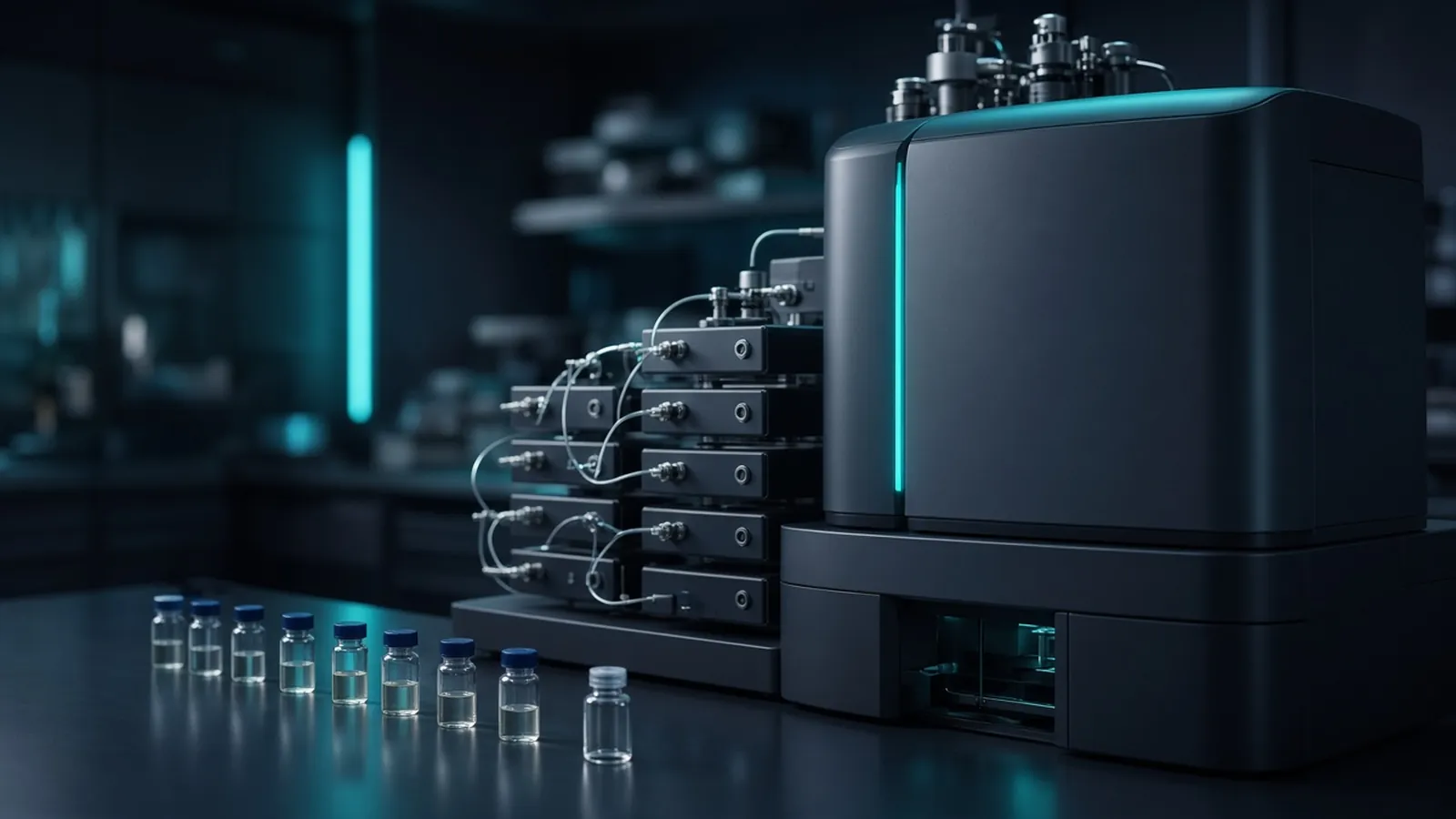
Preparing peptides for mass spectrometry is the work that decides whether the instrument sees your proteins or a cloud of detergent, salt and half-cut chains. Bottom-up experiments identify proteins from peptides, as the pillar page bottom-up proteomics in plain language explains. This guide stays at the decision level: which chemical step is for, how to tell it failed, and what the search will inherit. It is not a vendor microlitre table and not a clinical method.
What the digest has to deliver
The mass spectrometer, in the electrospray setups most proteomics labs use, wants peptides in a volatile solvent, free of non-volatile salt and of detergents that out-compete the analyte. The search engine wants peptide masses that match a simple rule: a named enzyme, cysteines in a known modified form, and only the extra modifications you deliberately allowed. Every shortcut in the tube becomes a wider search, and a wider search makes the false discovery rate work harder for a less specific list.
Start from a protein amount you measured, not from a guess. A vast excess of protein over enzyme leaves missed cleavages. A tiny amount disappears into tube walls and into keratin from the room. Record the estimate and the assay you used.
Reduction, alkylation and the cut
Disulfide bonds hold some chains in knots that proteases reach poorly. A reducing agent in the dithiothreitol or phosphine class opens those bonds. The alkylation step, classically an iodoacetamide-type reagent used with attention to light and time, prevents the bonds from closing and gives cysteine a defined mass. Quench or remove the leftover alkylating agent. An unquenched excess labels amines as well as cysteines, and those extra masses look like mysterious modifications.
Urea is a common denaturant because it unfolds proteins so the enzyme can work. Urea that is warm, old, or alkaline produces isocyanate, which carbamylates lysines and protein amino termini. A carbamylated lysine is a lysine trypsin may not cut. Keep urea steps cold and fresh, or choose a denaturant strategy that your method has already validated. Guanidinium chloride unfolds well and must be diluted or removed before trypsin, because the enzyme will not work in strong denaturant.
Trypsin is the default protease because it creates peptides with a basic residue at the end, which fragment in a helpful way. It is not the only choice. A protein with few lysines and arginines needs a different enzyme or a longer view of the sequence. Look at the sequence, on a resource such as UniProt, before you assume a trypsin digest will tile the chain. Digestion time and temperature belong to the enzyme lot you opened. An overnight digest is a habit, not a law. Over-digestion and autolysis of the enzyme itself add peptide noise.
Field guidance from the Human Proteome Organization is a useful reminder that identification standards assume the chemistry was controlled. Method write-ups on protocols.io and practical laboratory notes such as Addgene's protocol collection show how other groups document a digest. Adapt the controls, and keep your own volumes from the products you actually used.
Cleanup is part of the chemistry
Salts from the digest suppress electrospray and crust the source. A reversed-phase desalting step, in a cartridge or a stage tip, binds peptides, washes the salt away, and elutes them in an organic solvent that you then dry down. The resuspension solvent should be the one the liquid-chromatography method expects, usually a small share of acidified organic solvent in water. A sample resuspended in detergent-containing assay buffer is not ready, however carefully you digested it.
Sodium dodecyl sulfate is the frequent contaminant from lysates and from gel slices. It needs a removal method built for mass spectrometry: precipitation, a detergent-removal column, or a suspension-trapping style workflow are classes, each with losses. Coomassie and silver stains also ride along. A stained band is a localisation tool, not an injection solution.
Polymers from tubes, slips of plasticware, and some detergents produce repeating peaks that are obvious once you have seen them and invisible if you have not. If a scouting run shows a ladder of evenly spaced masses, stop and find the plastic or the soap. More gradient time will not turn polyethylene glycol into peptides.
Branch points
If the protein will not dissolve, do not digest the lump and call it a proteome. Change the denaturation, and record the loss. If a gel of the digest still shows the intact band, the enzyme did not work. Check the pH, the enzyme storage, and whether guanidine or detergent was still too high. If the scouting chromatogram is a series of polymer peaks, find the contamination before the long queue. If keratin dominates a sample that should be a bacterial lysate, the handling failed. Clean the bench and the tubes, and wear a coat you do not use for western blots of skin proteins.
A negative control digestion, with no sample protein, shows you the enzyme's self-peptides and the room's background. Those peptides will appear in the real samples too. Searching them as contaminants is more honest than pretending the background is zero.
| Observation | Likely meaning | Useful next step |
|---|---|---|
| Intact band remains after digestion | Protease did not cut | Check enzyme activity, denaturant dilution, and pH |
| Very high missed cleavages | Digest was incomplete or sites were blocked | Shorten the chemical story, or repeat with a fresh enzyme |
| Repeating mass peaks | Polymer or detergent | Trace tubes and soaps, then remake the sample |
| Almost only keratin | Handling background, or almost no analyte | Repeat preparation with cleaner handling and a known load |
| Empty chromatogram | Loss on cleanup, or ion suppression | Check the desalting elution and the resuspension |
How this changes the false discovery rate
The false discovery rate is applied after the search, using decoy sequences, as the pillar page describes. Preparation decides which peptides are in the vial and which modifications a truthful search must include. If carbamylation, over-alkylation and oxidation are all variable because the chemistry wandered, the search space grows. More candidates mean more chance matches at a given score. You can still set a one-percent threshold. The peptides that pass will be a thinner, stranger set, and protein inference will lean on fewer unique sequences. Controlling the chemistry is how you keep that threshold meaningful.
Safety, climate and handoff
Proteases, alkylating agents, acids and organic solvents need the chemical controls on their own safety sheets. Iodoacetamide-class reagents are toxic. Acetonitrile and acids used at the instrument are flammable or corrosive. Gel pieces add the acrylamide precautions of the gel workflow. Biological risk follows the source sample and your institutional rules. This guide does not authorise a diagnostic test.
In a hot room, urea left on the bench is more likely to break down. Chill the solutions the method requires, and do not assume a cupboard is cool because it is out of the sun. Power cuts during a vacuum dry-down are an inconvenience. Power cuts during a long acquisition are a failed file. Humidity and loose vial caps change the volume the autosampler picks up the next morning. When peptides travel, keep them cold or dry as agreed, and repeat a scouting injection if the cold chain lapsed.
What to send with an enquiry
Name the organism, the starting material, any detergent or denaturant, whether the proteins are already in a gel slice, the protease you expect, and whether the goal is identification or a quantitative comparison. The shotgun discovery proteomics reference, the protein identification reference, and the differential abundance reference are independent method pages for that conversation. Ask through the quote request whether a quotation is possible. Do not read them as a statement that EVRINTH runs the digest or operates the spectrometer. The useful specification is the cleanup and the search rules, not a catalogue adjective.
Prepare peptides that a mass spectrometer can actually read
- 01Remove the chemicals the ion source cannot tolerateKnow whether the sample still contains detergent, polymer, or a high salt. Plan a cleanup that removes them before anyone talks about injection volume.
- 02Reduce, alkylate, and digest with a recorded enzymeBreak disulfides, block the cysteines with the alkylating agent you chose, and digest with a stated protease. Keep urea cold if you use it, so you do not carbamylate the lysines you need trypsin to cut.
- 03Desalt into a mass-spectrometry solventA reversed-phase cleanup removes salts that suppress electrospray. Dry and resuspend only in the solvent the instrument method names.
- 04Judge the digest before a long runA small gel or a short scouting injection should show that intact protein has been cut and that the chromatogram is not a polymer ladder. A failed digest makes any later false-discovery threshold describe the wrong peptides.
Questions from the bench
Why alkylate cysteines before trypsin digestion?
Free cysteines can re-form disulfides and make peptides hard to see. Alkylation, often with an iodoacetamide-class reagent, caps those residues so the peptide mass is predictable. Too much reagent, or too long a reaction, also modifies other residues and complicates the search.
What does a high missed-cleavage rate tell me?
Trypsin did not finish. Causes include too much protein for the enzyme, an old enzyme, inaccessible sites, or lysines blocked by carbamylation or other modifications. The spectra can still match, and the protein inference gets harder and less complete.
How does preparation affect the false discovery rate?
The false discovery rate judges the search list you built. If the search must allow many unexpected modifications because the chemistry was dirty, more chance matches compete, and a given threshold keeps a less reliable set. Cleaner peptides let a simpler search do a stricter job.
Can I inject a gel slice that still smells of SDS?
Not usefully. Sodium dodecyl sulfate suppresses ionisation and contaminates columns. Remove it by a method validated for mass spectrometry, then desalt. A Coomassie-stained band still needs that cleanup.
References
Manufacturer names identify published method classes. Trademarks remain with their owners. Catalogue records on this site are independent references for enquiry. They are not a statement of inventory, distribution rights or a supply commitment. This page is educational. It is not medical advice, a diagnostic protocol or a biosafety approval.
Catalogue
Related products and categories
These links follow the subject of the article into published manufacturer references. A listing is a reference for an enquiry, not a statement of stock or distribution rights.
Continue in this cluster
Related reading
Bottom-up proteomics in plain languageBottom-up proteomics in plain language: proteins are digested to peptides, a mass spectrometer fragments them, and a database search names candidates.A glossary of proteomics termsA working glossary of proteomics terms, from precursor and PSM to razor peptide, FDR and batch effect, and how each term changes a pull-down or abundance claim.A protein ID is not a proof of functionA proteomics identification is a sequence match under a false discovery rate. Function still needs activity, phenotype, localisation or a perturbation, plus