
comparison
Antibody host species and secondary mismatch
How to match a secondary antibody to the primary host, subclass and label, and what a species mismatch does to the blank well.
- Author
- EVRINTH Editorial Team
- Published
- 8 October 2026
- Updated
- 8 October 2026
- Reading time
- 9 min
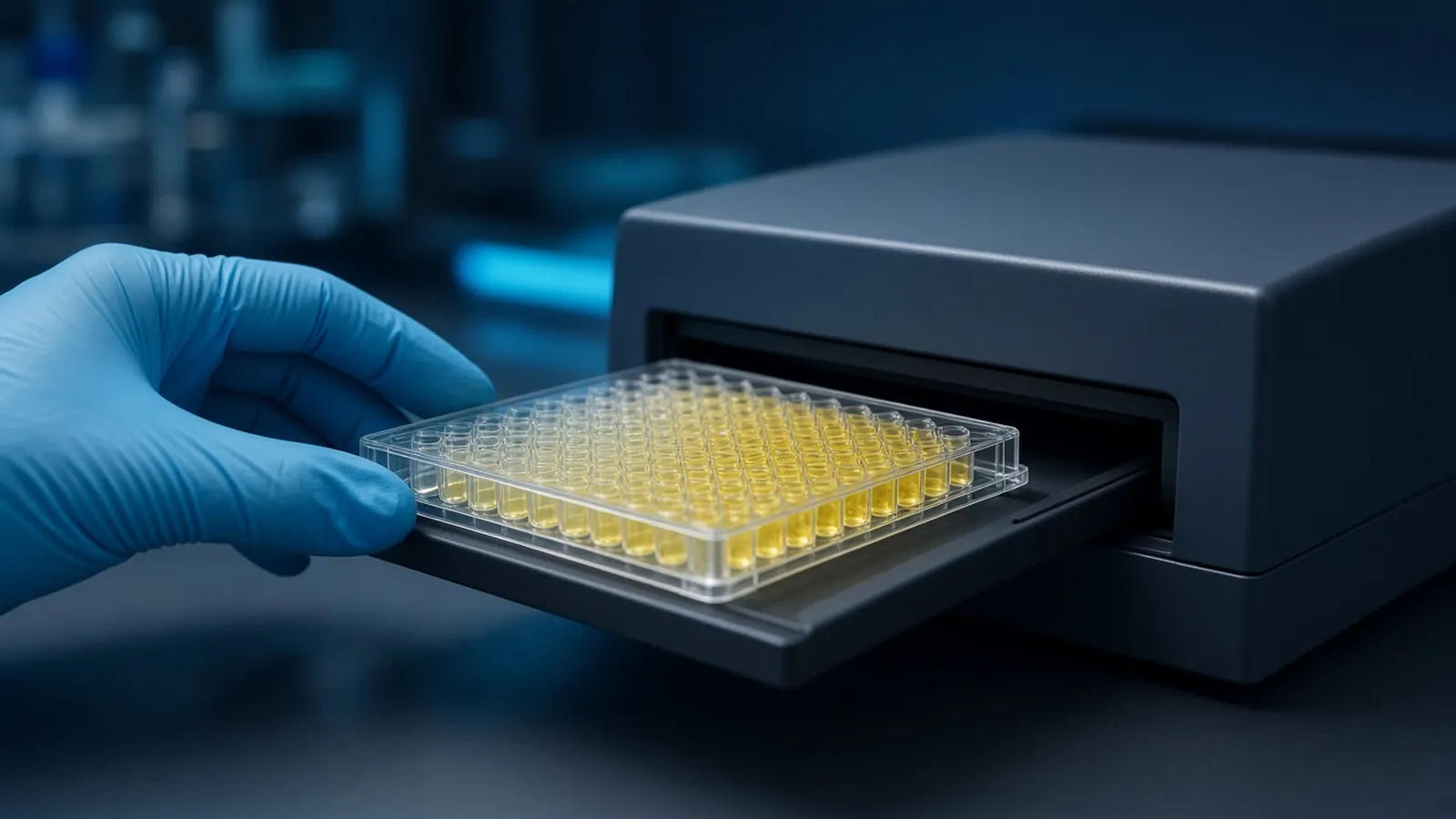
A secondary antibody is a reagent with a grammar. It must recognise the host species of the primary, it must recognise the class or chain you actually have, and its label must suit the reader. Goat anti-rabbit does not become a mouse detector because both tubes were in the same freezer box. This comparison is for someone writing a specification: primary host, secondary specificity, label class, and the species the secondary must not see in the sample. Plate formats that use these layers are in ELISA formats, controls and readout. How to test that the primary itself is specific is in choosing and checking a primary antibody. Trademarks on reagent names stay with their owners. Nothing here is a price or a sourcing promise.
The decision a specification has to freeze
Before a purchase request is written, the experiment already knows four facts. The primary antibody was made in a particular animal. It is a particular class, and if it is monoclonal it is a particular subclass. The sample comes from a species that has its own immunoglobulin. The readout is an enzyme, a fluorophore or a biotin handle. Leave any one of those out of the specification and the blank will teach it to you on the first plate, at the cost of the samples.
The comparison below is between a matched secondary and the classic mismatches. It is not a ranking of suppliers. Two matched reagents can still differ in cross-adsorption and in titre. The specification should say what must be true, and the first plate should test that statement.
The recognition rule, without decoration
A secondary antibody is an anti-immunoglobulin. The immunogen was IgG, or IgM, or a fragment, from one host species. Binding follows that immunogen. Rabbit primary, anti-rabbit secondary. Mouse primary, anti-mouse secondary. Rat, goat, chicken, guinea-pig and the rest follow the same rule. A chicken primary is often detected with anti-chicken IgY, because birds use IgY rather than IgG. Calling it "anti-IgG" on the paperwork is how that well goes blank.
Host of the secondary is chosen so it is not the host of the primary and not a species abundant in the sample. Goat anti-rabbit is common. Donkey secondaries are often chosen when several primaries from different hosts must be detected together, because a donkey can be immunised against several species and the resulting reagents can be absorbed against the others. That multi-label experiment needs a specification for each colour, plus a check that each secondary is quiet on the other primary.
The sample species is a third animal in the well. Mouse primary on mouse tissue, detected with anti-mouse, stains endogenous mouse immunoglobulin wherever plasma cells and serum have left it. The primary may be perfectly specific and the picture still wrong. The remedies are a different primary host, a method that avoids secondary antibody, or a tissue that truly lacks endogenous Ig. The same trap exists in a sandwich when the sample is serum of the same species as the capture or the detection reagent. Anti-mouse detection in a mouse serum sandwich can build signal on mouse Ig that has nothing to do with the analyte.
Subclass, chain and what the blot then shows
Monoclonal antibodies are one subclass. Mouse monoclonals are often IgG1, IgG2a, IgG2b or IgM. A secondary sold as anti-mouse IgG may be weak on IgG1, which is a known annoyance, or it may not see IgM at all. A secondary sold as anti-mouse IgG1 will miss an IgG2a clone. Write the subclass from the primary's sheet into the secondary line of the specification. If the sheet does not name a subclass, do not guess one in the purchase text. Ask, or buy a pan-IgG reagent and prove it on a known amount of that primary.
Heavy-chain and light-chain reactivity matter most on a reducing western blot and after immunoprecipitation. IgG falls apart into a heavy chain near 50 kilodaltons and a light chain near 25 kilodaltons, the exact apparent mass depending on the gel and the species. A secondary directed at Fc, the heavy-chain constant region, reports the heavy chain. A secondary directed at light chain reports kappa or lambda and therefore every class that uses that light chain. In a lysate blot that may be what you want. In a lane that contains the capture antibody, a light-chain band is often the reagent, not the antigen. Specify heavy-chain-only when that distinction is the point of the gel.
Label class is the last column of the specification. Horseradish peroxidase is the usual enzyme for colour ELISA and for chemiluminescent blots. Alkaline phosphatase is the alternative enzyme and it dislikes some phosphate buffers and some inhibitors. A fluorophore must name the dye closely enough that the filter set matches, and two dyes in one experiment must be spectrally separable. Biotin is not a signal. It is a handle for a later streptavidin enzyme, and it fails when the sample or the blocker already contains biotin. Pick one label class per readout. Do not plan to "see later" whether the plate reader can do fluorescence.
Sequence resources do not choose the secondary, but they stop you naming the wrong primary. Confirm the target accession on UniProt or NCBI Protein before you specify an antibody against it. Bench context for antibody handling sits in public notes such as the Addgene molecular biology reference and in method collections such as protocols.io.
What must not cross-react, written as a requirement
Anti-mouse reagents can see rat. Anti-goat reagents can see sheep and bovine immunoglobulin. Anti-rabbit reagents can pick up odd cross-reactions in other mammals if they were not absorbed. If the sample, the blocker or a second primary comes from one of those neighbours, the specification should say "cross-adsorbed against" that species, and the first experiment should include a well or a lane with that species' immunoglobulin and no primary. A quiet datasheet claim is a hypothesis. The well is the test.
| Primary host and class | Secondary that can see it | Classic mismatch | What the blank or the test well does |
|---|---|---|---|
| Rabbit IgG | Anti-rabbit IgG, labelled as required | Anti-mouse secondary used by habit | Signal stays at background. The primary bound and was never detected |
| Mouse IgG1 monoclonal | Anti-mouse IgG1, or a pan-mouse IgG shown to bind IgG1 | Anti-mouse IgM, or an IgG2a-only reagent | Blank-level signal on a true positive sample |
| Mouse IgM monoclonal | Anti-mouse IgM | Anti-mouse IgG | Same silence, often blamed on the antigen |
| Any primary on a blot after immunoprecipitation | Heavy-chain-specific secondary when the antigen sits near the light chain | Pan-Ig or light-chain secondary | A band at the light-chain mass that is the capture antibody |
| Mouse primary on mouse tissue or in mouse serum | A non-mouse primary host, or a method with no anti-mouse secondary | Anti-mouse secondary | Endogenous mouse Ig lights up. The no-primary control is already positive |
A bright blank with a structural cause
Mismatch toward silence is the easy failure. The positive control is blank and someone orders more antigen. Check the host line on both tubes before you change the biology.
Mismatch toward noise is the expensive failure. The secondary sees immunoglobulin that was already in the sample, the blocker or the other primary. The no-primary well is bright. Adding less analyte will not darken it. Either change the secondary to one adsorbed against that species, change the primary host, or change the blocker. A standard curve built on top of a bright blank can still be fitted. The concentrations will shift with the background and they will not mean what the axis says. Do not fit until the species stack is quiet.
A third failure is two primaries and two secondaries whose hosts cross. Rabbit and mouse is a classical pair because anti-rabbit and anti-mouse can be made not to see each other. Rabbit and goat is a worse pair if the anti-goat secondary was raised in rabbit, or if the anti-rabbit secondary sees goat. Draw the host of every antibody, including the host the secondary was raised in, as a small chain. Any loop in that chain is a blank that will light up.
Animal reagents and institutional rules
Secondary antibodies are animal immunoglobulin, often preserved with azide, sometimes labelled with an enzyme that has its own sensitisers. Fluorescent labels are a chemical class with their own waste rules. None of this is a diagnostic reagent because the hosts match. Research use stays inside the biosafety and chemical-safety rules of the institution. Human samples detected with anti-human secondary are still human samples. The specification does not lower the containment.
Cold chain, and a specification that survives the next lot
A secondary that thaws on a loading dock and then refreezes is a different reagent from the one specified, even if the host species on the label is perfect. Write the arrival condition into the receiving note: frozen, chilled, or warm, and whether the shipper still held cold packs. In a hot season that note matters more, because a short delay outdoors is enough to stress a conjugate. Aliquot after the first thaw if the sheet says repeated freeze-thaw costs titre. A power cut that warms the freezer is a lot event. Record it next to the vial.
When you raise a purchase specification, ask how the supplier documents the immunising species, the subclass reactivity, the adsorption against the sample species you named, and the label. Ask what a subsequent lot is compared against. You are asking for the contents of a document, not claiming that a certificate already exists in your file. Keep that wording tight so a later reader does not think the cross-adsorption was proved before you ran the no-primary well.
What the enquiry should pin down
State the primary host, the subclass if you know it, the sample species, the blocker species, and the label class the instrument can read. Say whether the experiment is a plate, a blot or a tissue section, because endogenous immunoglobulin is a different problem in each. Name any second primary that will share the sample. The reagents and chemicals catalogue is where related reagent classes can be browsed. The molecular biology pathway is the methods context. A matched secondary can be discussed through the quote request. Ask the reply to restate host, subclass and adsorption in words. A tube whose label and whose biology disagree will not be saved by a standard curve.
Questions from the bench
Does goat anti-rabbit detect a mouse primary antibody?
No. A secondary antibody is raised against immunoglobulin of a stated host species. Goat anti-rabbit IgG binds rabbit immunoglobulin. It does not bind a mouse primary, so the well stays at background even when the primary has found its target. Match the secondary to the animal that made the primary.
When does subclass matter more than species?
Species gets the secondary into the right animal. Subclass decides whether that secondary sees the particular monoclonal. An anti-mouse IgG1 reagent can miss a mouse IgM or a mouse IgG2a clone. If the datasheet of the primary names a subclass, the secondary specification has to name it too.
Why does a light-chain secondary change a blot?
IgG heavy chain and light chain separate on a reducing blot and sit at different apparent masses. A secondary that sees only heavy chain reports the larger chain. A secondary that sees light chain, or that sees both, also reports the smaller chain. In an immunoprecipitation lane that smaller band can be the capture antibody's own light chain rather than the antigen.
Are enzyme and fluorophore labels interchangeable?
They answer different readers. Horseradish peroxidase and alkaline phosphatase need a substrate and a plate reader or a blot imager. A fluorophore needs the right excitation and emission and suffers from overlap with other dyes. Changing the label class changes the instrument and the background chemistry, not only the colour of the tube cap.
References
Manufacturer names identify published method classes. Trademarks remain with their owners. Catalogue records on this site are independent references for enquiry. They are not a statement of inventory, distribution rights or a supply commitment. This page is educational. It is not medical advice, a diagnostic protocol or a biosafety approval.
Catalogue
Related products and categories
These links follow the subject of the article into published manufacturer references. A listing is a reference for an enquiry, not a statement of stock or distribution rights.
Continue in this cluster
Related reading
ELISA formats controls and readoutELISA formats, controls and readout: how direct, sandwich and competitive assays turn a binding event into a number that stays inside the curve. Choosing and checking a primary antibodyHow to choose and check a primary antibody: host, clonality, application claims, and controls that show the signal is the intended target.Isotype controls are not a full answerWhat an isotype control actually measures, which sticky artefacts it can rule out, and why it cannot replace a knockout or a secondary-only well.