
guide
Choosing and checking a primary antibody
How to choose and check a primary antibody: host, clonality, application claims, and controls that show the signal is the intended target.
- Author
- EVRINTH Editorial Team
- Published
- 8 October 2026
- Updated
- 8 October 2026
- Reading time
- 7 min
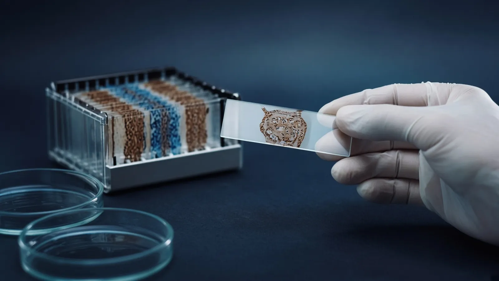
Choosing and checking a primary antibody is an experiment, not a catalogue filter. The reagent binds an epitope. Your sample contains thousands of other surfaces. A datasheet photograph is evidence about the supplier's sample, under the supplier's conditions. Your job is to show that the band, the stain, or the ELISA signal in your hands depends on the target you named. Plate formats and curves are the companion pillar ELISA formats, controls and readout. This guide is for research use. It is not a diagnostic validation.
What to read before you buy
Start with the target as a sequence, not as a gene symbol that several species share. UniProt and Ensembl let you check whether the immunogen peptide exists in the species and the isoform you study. An antibody raised against a human peptide can fail on a mouse protein that differs at that stretch, or it can cross-react with a related protein that kept the stretch. Write the accession you checked.
Host species has to work with the detection system. A mouse primary antibody on mouse tissue, detected with an anti-mouse secondary, will paint endogenous immunoglobulin unless you have a method that avoids that. The host should also differ from the capture or secondary reagents in a sandwich assay so the enzymes bind the right layer.
Clonality is a trade. A monoclonal antibody is one epitope, one reproducible binding site, and a risk that the epitope is modified, cleaved, or shared. A polyclonal antibody is a mixture that can survive partial epitope loss and can change when a new animal is immunised. Recombinant antibodies are a class that can be defined by sequence, which makes lot changes easier to discuss. None of these labels replaces a gel or a slide.
Application claims are narrow. An antibody that works on a denatured blot may fail on a fixed tissue section, because fixation hides or destroys epitopes. The reverse happens too. The photograph of a stained slide is a reminder that tissue staining has its own controls: morphology, a negative tissue, and a no-primary slide. Do not transfer a dilution from a blot to a section and call it the same assay.
How to check it with samples you understand
A positive control expresses the target for a reason you can explain: a lysate from a cell line known to express it, a recombinant protein, or a tissue where independent evidence says the protein is present. A negative control lacks it for a reason you can explain. A knockout or knockdown is the strongest genetic control. Editing resources such as the Addgene CRISPR guide are a public orientation to how research laboratories build that kind of cell, not a protocol you can skip validating. If you do not have a knockout, a cell or tissue that genuinely does not express the transcript is a weaker but still useful negative, provided you did not choose it only because the antibody was blank there.
The no-primary control uses blocking and the secondary detection exactly as in the real samples, without the primary antibody. Signal there belongs to the secondary, to endogenous enzyme, or to sticky tissue. Blocking buffers, discussed for plates in the ELISA article, matter on blots and on slides for the same reason. Change the blocker when the no-primary slide is already stained. Do not only shorten the development.
An orthogonal check uses a different physical principle. A band that appears with two antibodies against different epitopes, or a band that matches a mass-spectrometric identification of that slice, is stronger than a second aliquot of the same antibody. Peptide pre-absorption can reduce a specific signal and can also reduce a non-specific signal if the non-specific binder likes that peptide. Treat it as one clue.
Dilution series tell you where the specific signal remains and the extra bands fade, or where everything fades together. The second pattern is not specificity. Record the lot number. A new lot of a polyclonal antibody is a new reagent.
Where a standard curve belongs
Once the signal depends on the target, quantification can be discussed. On a blot, densitometry needs a linear range and a loading control that is stable in your treatment. On an ELISA, a standard curve, built as the format article describes, converts signal to concentration inside that curve. Spike recovery and dilution parallelism still apply. A curve does not repair a no-primary well that is already bright, and blocking does not repair an antibody that binds the wrong protein in both the standard and the sample.
| Check | Supports | Does not support |
|---|---|---|
| Immunogen present in your species | The epitope might exist | That the reagent is specific in lysate or tissue |
| Band at the expected mass | Size is consistent with the target | That no other protein co-migrates |
| Signal lost in a knockout | Dependence on that gene in this assay | Every extra band on a different tissue |
| No-primary slide or lane is quiet | Detection reagents are not the stain | That the primary antibody is selective |
| Standard curve and spike recovery | The range and the matrix, after specificity | Specificity itself |
Failure modes
A single band at the "right" size in every sample, including the negative, is a cross-reactive protein or a loading artefact. Extra bands that vanish in the knockout may be true fragments. Extra bands that remain are not. Tissue edges that stain only at the margin are often drying artefacts. Nuclear stain with an antibody against a secreted protein is a reason to stop and look at the secondary and the antigen retrieval, not a reason to rewrite the cell biology.
Isotype controls match the antibody class and do not match the specificity. They help with some flow-cytometry backgrounds and do not prove a band on a blot. Trusting a dilution posted for a different tissue is a common way to over-stain. Find the dilution in your system.
Safety, storage and the enquiry
Preservatives such as sodium azide are toxic and also inhibit horseradish peroxidase if they are carried into a detection step that uses that enzyme. Fluorophores fade. Fixed tissue and human samples follow your biosafety rules. This page does not interpret a stain as a diagnosis.
Antibodies are cold-chain reagents. Record the arrival temperature. Repeated freeze-thaw cycles of a small vial kill some clones and leave others. Aliquot on arrival if the datasheet says to freeze, and keep the lot identity on the aliquot. In a humid cold room, labels fall off. A power cut that warms a refrigerator is a reason to re-run the positive and negative controls before a large experiment. Do not assume a vial that stayed "cool to the touch" is unchanged.
For a sourcing note, name the target accession, the species of the sample, the application, the host species you can detect, any fixation, and the positive and negative samples you will use to check the lot. Start from the reagents and chemicals catalogue and the molecular biology pathway. Use the quote request to ask whether a quotation is possible. A family name on a product line is not a promise of a particular clone, and this site does not claim to hold that vial or to validate it for you. Ask for the immunogen and the application data with the offer.
Method notes on protocols.io are a reasonable place to see how other groups document antibody controls. Keep your own images with the lot numbers attached.
Accept a primary antibody only after a local check
- 01Match the reagent to the sample and the applicationConfirm species reactivity, the immunogen, the host species, and that the supplier tested the application you will use. A western blot claim is not an immunohistochemistry claim.
- 02Run a positive and a negative sampleInclude a sample that should contain the target and one that should not, such as a knockout or a cell line that does not express it. Similar staining in both is not specificity.
- 03Run the secondary aloneA no-primary control shows what the detection reagents and the blocking do by themselves. Subtracting that image is not a substitute for a quiet control.
- 04Retire the lot when the check failsPolyclonal lots differ. If the band or the stain does not depend on the target, stop using that lot. A standard curve comes only after specificity is believable.
Questions from the bench
Is a monoclonal antibody automatically more specific than a polyclonal?
It is specific for one epitope, which can be an advantage and can also be a shared epitope. A polyclonal reagent sees several epitopes and can be more tolerant of a damaged antigen, with a higher chance of extra bands. You still have to test both in your samples.
Does a band at the right molecular weight validate the antibody?
It is one supportive observation. Other proteins share that mass, and a specific antibody can still show extra bands from breakdown or cross-reactivity. A negative sample that lacks the target, or an orthogonal method, is the check that makes the mass meaningful.
Where does a standard curve fit in antibody checking?
After you believe the signal is the analyte. In an ELISA, the curve then shows the range where the antibody pair reports concentration. A beautiful curve from a cross-reactive antibody is a precise measurement of the wrong thing.
Can I use a research antibody result as a pathology report?
No. Immunohistochemistry and immunoassays used for diagnosis are validated for that purpose under a quality system. A research stain supports a research claim about the samples you controlled.
References
Manufacturer names identify published method classes. Trademarks remain with their owners. Catalogue records on this site are independent references for enquiry. They are not a statement of inventory, distribution rights or a supply commitment. This page is educational. It is not medical advice, a diagnostic protocol or a biosafety approval.
Catalogue
Related products and categories
These links follow the subject of the article into published manufacturer references. A listing is a reference for an enquiry, not a statement of stock or distribution rights.
Continue in this cluster
Related reading
 ELISA formats controls and readoutELISA formats, controls and readout: how direct, sandwich and competitive assays turn a binding event into a number that stays inside the curve.A glossary of immunoassay termsAnalyte, capture, conjugate, blank, matrix, spike recovery and lot, defined so a datasheet can be shortlisted before a reagent is ordered.Antibody host species and secondary mismatchHow to match a secondary antibody to the primary host, subclass and label, and what a species mismatch does to the blank well.