
Pillar guide
ELISA formats controls and readout
ELISA formats, controls and readout: how direct, sandwich and competitive assays turn a binding event into a number that stays inside the curve.
- Author
- EVRINTH Editorial Team
- Published
- 8 October 2026
- Updated
- 8 October 2026
- Reading time
- 8 min
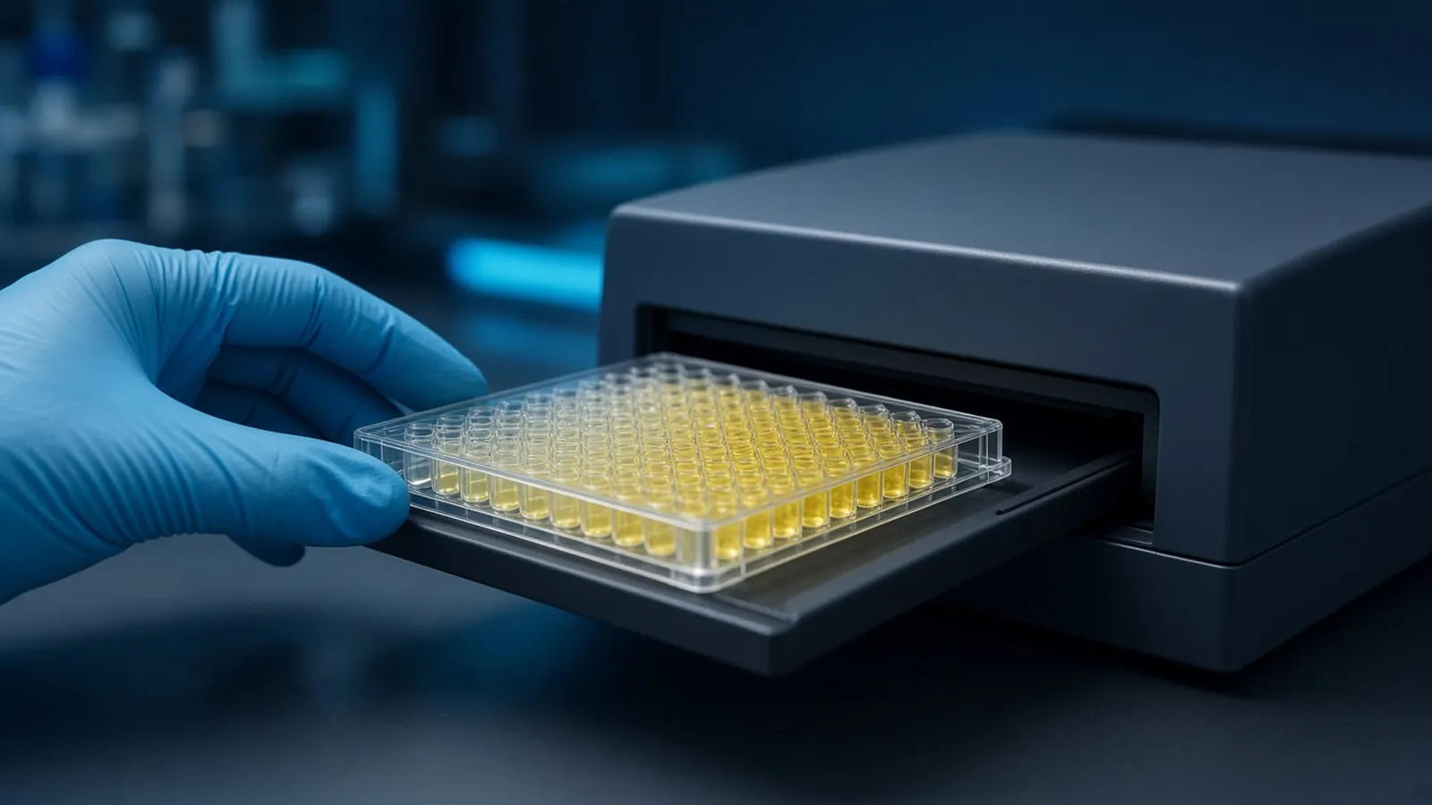
ELISA formats, controls and readout are the three decisions that turn an antibody binding event into a number you can say out loud. The plate does not know your hypothesis. It reports enzyme activity. You earn the right to call that activity a concentration by choosing a format that matches the analyte, by running controls that would have caught a sticky well, and by reading only inside a standard curve. Choosing the primary antibody itself is the companion guide choosing and checking a primary antibody. This page is a research explainer, not a diagnostic instruction.
Which format matches the analyte
In a direct assay the antigen is coated on the plastic and a labelled primary antibody binds it. It is simple and it needs a relatively pure antigen. An indirect assay uses an unlabelled primary antibody and a labelled secondary antibody, which adds signal and also adds a place for the secondary to stick. A sandwich assay coats a capture antibody, lets the analyte bind from a mixture, and detects it with a second antibody on a different epitope. That is the usual choice for a protein in serum, lysate, or culture medium, because the capture step supplies specificity the plastic cannot.
A competitive assay incubates the sample with a limited amount of antibody, or competes the sample against a labelled antigen. Signal falls as the analyte rises. Many small molecules are measured this way because they have only one epitope and cannot form a sandwich. Read the direction of the curve before you rank samples. A darker well is not automatically "more analyte".
The antibodies have to fit the format. A sandwich needs two clones, or a polyclonal pair, that can bind at once. Check that claim with a control antigen, not with hope. Sequence context for the target, including domains a capture peptide might have missed, belongs on a page such as UniProt.
How binding becomes a number
Most research plates use an enzyme conjugate, often horseradish peroxidase or alkaline phosphatase, and a substrate that becomes coloured or luminescent. The microplate reader in the photograph is the measurement, and it is only as meaningful as the wells you built. For a colorimetric substrate, the wavelength and any stop reagent belong to that substrate. Copy them from the reagent you opened. A kinetic read and an endpoint read are different experiments. Say which one you did.
The standard curve is a set of known concentrations taken through the same protocol. Fit a line only when the chemistry is actually linear. Many ELISA curves are sigmoid across a wide range, and a four-parameter fit is a common description. Choose the fit because it matches the curve, and lock that choice before you interpret outliers. Concentrations below the lowest standard or above the highest are outside the assay. Dilute or concentrate, or report them as out of range.
Matrix is part of the curve. A standard diluted in buffer can sit on a different line from the same standard diluted in serum. Spike a known amount into the real sample matrix and ask what fraction you recover. Poor recovery means the number is a rank at best. Dilutional parallelism, where a diluted sample falls on the same line, is the check that you are measuring the same thing at different doses.
Blocking, washes and the primary antibody
Blocking is a protein or detergent solution that covers bare plastic after the capture step. Without it, the detection antibody binds the plate and every well lights up. The no-analyte well is the proof. If that well matches your low samples, you do not have a low sample. You have a background.
Washes remove unbound reagent. Too few washes raise the background. Too many, or a harsh buffer, can strip a weak interaction. Record the buffer and the count. The primary antibody's dilution is empirical. A checkerboard of capture and detection dilutions, run once when the assay is set up, saves every later plate. The antibody guide explains how to decide the reagent is specific before you invest in that checkerboard.
Method collections such as protocols.io show how other laboratories structure a plate map. Keep your map in the same file as the absorbance values. A plate map reconstructed the next day is how standards and samples trade names.
Controls worth the wells
A substrate blank contains substrate and stop, without enzyme. It catches a substrate that has already gone off. A zero standard contains matrix and no added analyte. It catches background and contaminated diluent. A positive control of known concentration, ideally a lot you kept for this purpose, catches a dead enzyme or a forgotten antibody. A well with no capture antibody, or no primary antibody, catches reagents that bind plastic or the sample directly.
Replicates show pipetting. They do not create biological replication. If you plated one culture three times, you have one biological result.
| Well | What a clean result supports | If it fails |
|---|---|---|
| Substrate blank | The substrate was quiet before enzyme | Discard the substrate. Do not subtract a large blank and continue |
| Zero standard | Blocking and antibodies are not painting the matrix | Change blocker, wash, or antibody before you trust lows |
| Standard curve | Response spans the concentrations you claim | Report out-of-range samples as out of range |
| Spike recovery | The matrix is not hiding or inflating the analyte | Quantify only after the matrix is handled, or report a rank |
| No-primary control | Colour depends on the primary antibody | The secondary or the enzyme system is the signal |
Failure modes
The hook effect, already noted, is the dramatic one. Edge effects are the quiet one. Outer wells evaporate faster, concentrate the reagent, and drift. If the plate map puts all the controls in the centre and all the treated samples on the edge, the map is the result. Rotate layouts, or avoid edges, and seal during incubations. Temperature gradients in a cold room or a sunny bench do the same thing.
A biotin-avidin amplification system fails in samples rich in biotin, including some milk blockers. A phospho-specific detection fails in milk that contains phosphoproteins. Cross-reactivity of the primary antibody is a reagent problem, taken up in the antibody article. An expired substrate turns the whole plate yellow, including the blank. That plate is not "high expression".
Safety and research limits
Antibodies and standards may contain azide or other preservatives. Stop solutions are often acid. Substrates can be irritants. Human serum and culture samples that might contain pathogens follow institutional biosafety rules. The WHO Laboratory Biosafety Manual and the CDC BMBL are public references for that institutional decision. They are not a permit, and this article is not a diagnostic approval.
Heat, humidity and the reader
Colour development is a timed enzyme reaction. A hot afternoon shortens it. Keep the development in the temperature window the substrate states, and use the same timer for every plate you will compare. Humidity and a loose lid change edge wells first. If a power cut stops the reader after development, the colour may still be on the plate. Read it promptly and record the delay. A stopped reaction is more patient than a kinetic one. Do not leave a developed plate in direct sun while you find a spare reader.
What to put in an enquiry
Name the analyte, the sample matrix, the species, whether you need a sandwich or a competitive format, the concentration range you care about, and any biotin or phospho constraint. The reagents and chemicals catalogue is the sourcing start for buffers, plates and related reagent classes. The molecular biology pathway is the wider methods context. Use the quote request to ask whether a quotation is possible. Do not treat a catalogue line as a coated plate that is already validated for your matrix, and do not read this site as operating a diagnostic ELISA service. Ask for the antibody pair and the curve range in the reply.
Choose an ELISA format and the controls that make the number real
- 01Match the format to the analyte and the sampleUse a sandwich assay for a protein in a complex mixture when you have two antibodies on different epitopes. Use a competitive format for many small molecules. A direct coat of pure antigen answers a narrower question.
- 02Block, and prove the block with a no-analyte wellBlocking reduces sticky signal. A well with no analyte, carried through every other step, shows whether the block and the antibodies are quiet enough to read.
- 03Build a standard curve in an honest matrixDilute a known standard in the same kind of matrix as the samples when you can. Read concentrations only between the standards, with the curve fit you stated before looking at the samples.
- 04Check hook effect and edge wellsDilute high samples to see whether the signal falls as it should. Treat edge wells with suspicion if evaporation or temperature made them outliers.
Questions from the bench
Why did a very concentrated sample read lower than a dilute one?
Sandwich assays can show a hook effect. Excess analyte occupies capture and detection antibodies separately, so the sandwich never forms and the signal drops. Dilute the sample. If the dilute well reads higher, the neat well was past the assay, not below it.
Does a yellow well prove the analyte is present?
A colour change shows that enzyme met substrate. It is evidence of analyte only when the blank, the zero standard, and a control without primary antibody are quiet, and the sample sits on a valid curve. Sticky plates and cross-reactive antibodies also turn yellow.
What is blocking actually doing?
Blocking occupies plastic that would otherwise bind detection reagents. Albumin, casein, milk and commercial blockers are classes with different failures. Milk can interfere with biotin or phospho-specific systems. The right blocker is the one your controls accept.
Is a research ELISA a diagnostic test?
Not by default. Diagnostic immunoassays are validated for a stated specimen and a stated decision. A research plate supports the comparison you designed. It does not become clinical because the wavelength was 450 nanometres.
References
Manufacturer names identify published method classes. Trademarks remain with their owners. Catalogue records on this site are independent references for enquiry. They are not a statement of inventory, distribution rights or a supply commitment. This page is educational. It is not medical advice, a diagnostic protocol or a biosafety approval.
Catalogue
Related products and categories
These links follow the subject of the article into published manufacturer references. A listing is a reference for an enquiry, not a statement of stock or distribution rights.
Continue in this cluster
Related reading
 Choosing and checking a primary antibodyHow to choose and check a primary antibody: host, clonality, application claims, and controls that show the signal is the intended target.A glossary of immunoassay termsAnalyte, capture, conjugate, blank, matrix, spike recovery and lot, defined so a datasheet can be shortlisted before a reagent is ordered.Antibody host species and secondary mismatchHow to match a secondary antibody to the primary host, subclass and label, and what a species mismatch does to the blank well.