
guide
Bradford BCA and absorbance at 280 nm
Bradford, BCA and absorbance at 280 nm report different chemistries. Pick the assay your buffer allows and name the standard beside the number.
- Author
- EVRINTH Editorial Team
- Published
- 8 October 2026
- Updated
- 8 October 2026
- Reading time
- 8 min
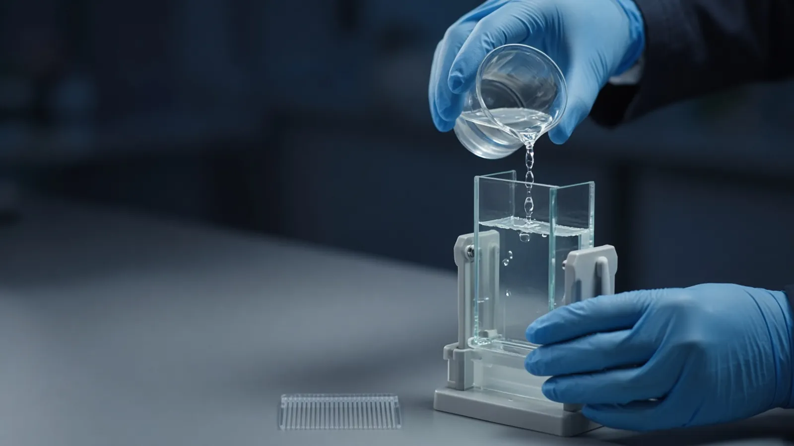
A number written on a tube of protein is only as good as the chemistry that produced it. Bradford dye binding, a bicinchoninic acid copper assay, and absorbance at 280 nm can all be called protein quantification, and they do not answer the same question. One is a colour from a dye, one is a colour from reduced copper, and one is ultraviolet light absorbed by a few side chains. The decision this page supports is which of those readings you are allowed to use for the buffer in front of you, and what you must write beside the number so the next person can repeat it. The companion on how laboratories estimate protein concentration sets the wider frame. A gel, covered in reading a protein gel, still has to show whether that mass is one polypeptide.
What each reading is actually made of
The Bradford assay, introduced as a protein-dye method by Bradford in 1976, uses Coomassie dye. Free dye and protein-bound dye absorb light differently, so the well changes colour as protein binds. Binding is not a fair census of peptide bonds. Arginine, and to a lesser extent lysine, histidine and some aromatic residues, contribute strongly. A basic protein and a small acidic protein of equal mass can give unequal colour. Immunoglobulin and albumin are a practical pair that often disagree. That protein-to-protein variation is a property of the chemistry, not a pipetting error.
Detergent is the other Bradford problem. Sodium dodecyl sulfate, Triton-type detergents and some other surfactants disturb the dye equilibrium. The direction and the size of the error depend on the formulation and the concentration. A lysate prepared for electrophoresis, with SDS already in it, is a poor candidate for a classical Bradford reading. Some commercial versions raise the detergent tolerance. That claim belongs to the bottle you bought. It does not transfer to a different Coomassie recipe.
The BCA assay, described by Smith and colleagues in 1985, is a copper method. In alkaline solution, peptide bonds and some side chains reduce Cu2+ to Cu1+. Bicinchoninic acid then forms a coloured complex with the reduced copper. Because the peptide backbone contributes, the protein-to-protein spread is often narrower than with Coomassie, though it does not disappear. BCA formulations are widely chosen when the sample must contain detergent, because many detergents that ruin a Bradford plate are tolerated here. The trade is reductant. Dithiothreitol, beta-mercaptoethanol and related reducing agents reduce copper themselves, so they invent colour. Chelators such as EDTA can pull copper out of the reaction. A high concentration of a reducing sample buffer is a reason to change the assay or to remove the reductant first, not a reason to subtract a guessed blank.
Absorbance at 280 nm does not add a dye or a metal. Tryptophan, tyrosine and cystine account for almost all of the near-ultraviolet absorption of a typical polypeptide. A pure protein, a 1 cm path, and an extinction coefficient that matches the construct convert absorbance into concentration. Nucleic acids absorb strongly in the same region and inflate the number. The ratio of absorbance at 260 nm to absorbance at 280 nm is the usual warning: protein-rich samples sit lower than nucleic-acid-rich samples. Imidazole and some buffers also absorb. A water blank under a sample dissolved in urea or a coloured component is not a blank. The assumptions packed into a calculated coefficient are set out in what an extinction coefficient assumes.
The standard is part of the result
Bovine serum albumin is common because it is available and usually well behaved in these assays. It is not your protein unless you are measuring albumin. A Bradford number read against albumin and reported as if it were milligrams of your enzyme hides the dye-binding difference. Say albumin equivalents when that is the scale you used. If you later switch to a standard made from the purified protein itself, expect the number to move. Both readings can be careful. They are not interchangeable in a table.
For a pure protein with a known coefficient, A280 avoids that translation step. For a crude lysate you will load on a gel, an albumin-equivalent dye or copper assay is an operational load, not a molarity of the enzyme you care about. Inclusion-body material dissolved in urea or guanidine needs an assay the denaturant does not break. High denaturant often pushes people toward A280 with a matched blank, once a coefficient exists. Check the interference notes for the reagent generation you have. Public method notes on protocols.io are useful for seeing where laboratories place these assays. They are not a substitute for the insert in the box.
A path with branch points
Start from the additives, not from the assay the bench next door prefers. If the sample contains SDS or another strong detergent and little reductant, a BCA-type copper assay is usually the candidate to test. If the sample is a simple salt buffer and you can accept protein-to-protein variation, Bradford is often the faster colour. If the sample is a single known polypeptide and nucleic acid is low, measure A280 and record the coefficient and the path length.
Then look at the blank. A blank that already looks as blue or as purple as the standards means the buffer is creating signal. Stop and identify the additive. Do not subtract an overflowing blank and call the remainder a concentration. If standards are flat, the reagent may be old, the incubation may be wrong, or a chelator may have removed the copper. Follow the manufacturer protocol for time and temperature. Do not invent a microlitre table from memory or from a different kit.
If the unknown is darker than the top standard, dilute it in the same buffer and read again. A value past the curve is a prompt to dilute. If Bradford and A280 disagree on a pure protein, believe the disagreement. It is information about composition, dye response, or a contaminant. Run the sample on a gel before you average the two numbers into a false compromise.
| Assay | Signal comes from | Often troubled by | Fits best when |
|---|---|---|---|
| Bradford (Coomassie) | Dye binding, strong for some residues | Many detergents, large protein-to-protein gaps | Simple buffers, and a named standard is acceptable |
| BCA (copper) | Cu1+ complex after alkaline reduction | Reducing agents, some chelators | Detergent is present and reductant is absent |
| A280 | Trp, Tyr and cystine | Nucleic acids, cofactors, absorbing buffers | One polypeptide and a matching coefficient |
Readings that look precise and are not
A spectrophotometer can print three decimal places for a cloudy sample. Scatter is not absorption by tryptophan. Spin or filter first, and read the cleared liquid, as the aggregation note in this cluster describes. A plate reader with a short path does not automatically match a 1 cm coefficient. Either use the instrument's path-length correction as the manufacturer defines it, or stay with a relative standard curve and say so.
Old Coomassie reagent and a copper reagent that has sat warm can both drift. A standard curve that no longer resembles last month's curve is a failed control. Make a fresh curve. Do not apply last month's slope to today's wells. If duplicates scatter, suspect mixing, bubbles, or a sample that is still settling, before you suspect the biology.
Handling, research use and the room
Coomassie reagent, alkaline copper reagent and the acids used around them are chemical hazards. Follow the safety note for the formulation you have. This page is a research explainer. It is not a clinical protein method and not biosafety approval. Your institution decides containment for the lysate.
In a hot, humid laboratory, "room temperature incubation" is not a temperature from a temperate kit insert. Keep the incubation in the range the protocol states, and do not leave a dye reagent in direct sun on a bench. A spectrophotometer that rebooted after a power cut needs a lamp warm-up and a standard check before a precious dilution series is trusted. Weigh hygroscopic standards with the same care you would give any other salt that takes up water. Record the assay, the standard, the dilution and the buffer. Those four words stop a later argument about whose number was right.
What to put in a reagent enquiry
Name the sample matrix first: lysate, elution, inclusion-body solubilisation, or a polished pool. Name the detergents and reductants at the concentrations you actually use. Say whether you need albumin equivalents for gel loading or a molarity tied to a coefficient. Reagents and chemicals is the catalogue family for assay components and buffer salts. A quote can carry that matrix description. The custom protein expression and purification page is an enquiry reference if the quantification sits inside a larger expression question. It is a place to discuss a method. It is not a statement that a purification is already being run.
Choose a protein assay the buffer will allow
- 01Write down every additive in the tubeList detergent, reductant, chelator, urea or guanidine, imidazole and any coloured component. The list is the filter that removes assays before you open a plate.
- 02Match one chemistry to that listUse dye binding when detergent is absent and protein-to-protein variation is acceptable. Use a copper assay when detergent is required and reductant is not. Use 280 nm when the polypeptide is pure enough and an extinction coefficient fits the construct.
- 03Blank and standard in the same matrixThe blank contains the buffer without the protein you claim to measure. The standard is either the protein itself or a named substitute such as bovine serum albumin, and the reported number says which one you used.
- 04Keep the reading inside the curveDilute until the signal sits among the standards. Follow the manufacturer protocol for incubation and wavelength, and attach the assay name, standard and dilution to the value you write down.
Questions from the bench
Why do Bradford and BCA disagree on the same lysate?
They detect different chemistry. Bradford dye binding depends strongly on the amino-acid composition, while a BCA assay follows copper reduced by the peptide backbone and some side chains. Two internally consistent curves can still produce two different albumin-equivalent numbers.
Does absorbance at 280 nm need a standard curve?
For a pure polypeptide with a suitable extinction coefficient and a known path length, the Beer-Lambert relationship replaces a protein standard. You still need a matched buffer blank. If nucleic acid or a cofactor also absorbs, the reading is no longer a concentration of that polypeptide alone.
Which standard should I choose, albumin or my own protein?
Use the protein itself when you have a trustworthy pure sample and the assay responds to it. Use bovine serum albumin when you need a shared operational scale, and call the result albumin equivalents. Switching the standard changes the number even when the colour in the well does not.
Can I copy microlitre volumes from a different kit insert?
No. Formulations differ in dye strength, copper reagent and the range they cover. Follow the protocol that belongs to the bottle in your hand, and treat a published table from another supplier as background reading rather than a recipe.
References
Manufacturer names identify published method classes. Trademarks remain with their owners. Catalogue records on this site are independent references for enquiry. They are not a statement of inventory, distribution rights or a supply commitment. This page is educational. It is not medical advice, a diagnostic protocol or a biosafety approval.
Catalogue
Related products and categories
These links follow the subject of the article into published manufacturer references. A listing is a reference for an enquiry, not a statement of stock or distribution rights.
Continue in this cluster
Related reading
Reading a protein gelHow to read a protein gel: what SDS-PAGE bands, smears, ladders and loading differences can support, and what they cannot identify. How laboratories estimate protein concentrationHow laboratories estimate protein concentration with absorbance, dye-binding and copper assays, and why the number depends on the standard.What an extinction coefficient assumesA composition-based extinction coefficient is a conversion with conditions attached. Tags, cofactors and nucleic acid change the number you may report.