
guide
How laboratories estimate protein concentration
How laboratories estimate protein concentration with absorbance, dye-binding and copper assays, and why the number depends on the standard.
- Author
- EVRINTH Editorial Team
- Published
- 8 October 2026
- Updated
- 8 October 2026
- Reading time
- 7 min
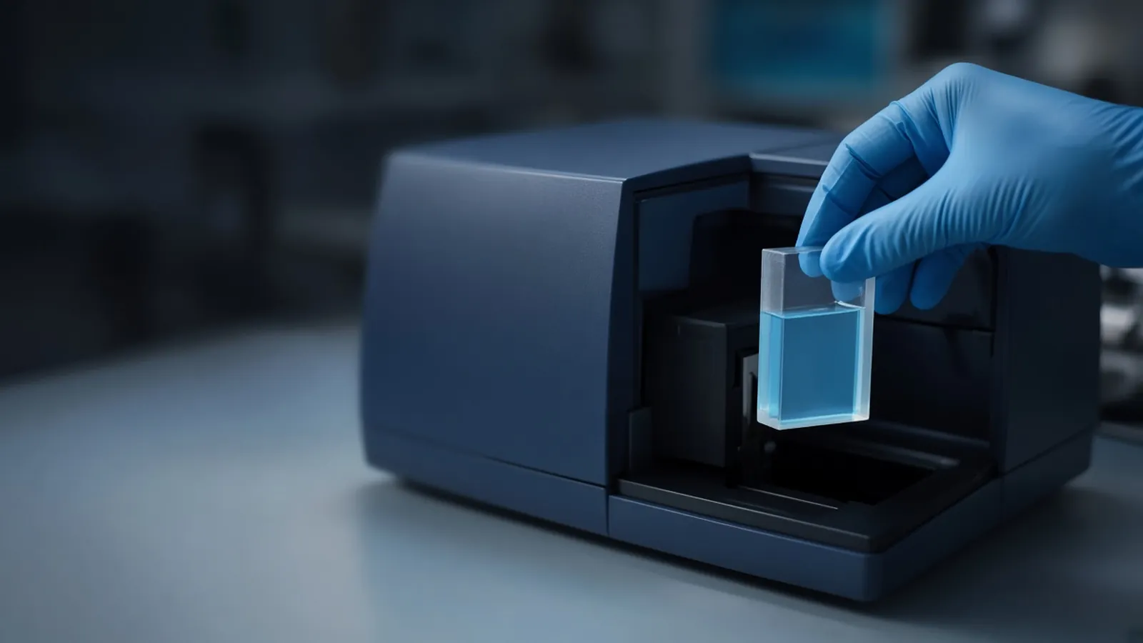
How laboratories estimate protein concentration is a choice of chemistry, a standard, and a blank. The number is an estimate of signal converted through those choices. It becomes useful when the next person can repeat it. It becomes misleading when the buffer, the standard, or the instrument is left off the label. This guide sits beside reading a protein gel. The gel asks which polypeptides are present. The assay asks how much signal the solution produces. Neither one replaces the other.
Three chemistries, three different "amounts"
Absorbance at 280 nm uses the ultraviolet absorption of tryptophan, tyrosine, and cystine. For a pure protein, a path length, and an extinction coefficient, the Beer-Lambert relationship converts absorbance into concentration. Many database entries, including those on UniProt, list a predicted extinction coefficient for the unmodified chain. Use the coefficient that matches your construct, including tags if they contain aromatic residues. Nucleic acids absorb strongly nearby, so a protein that still contains DNA will look more concentrated than it is. A ratio of absorbance at 260 nm to 280 nm is a warning light for that problem.
A Bradford assay is the dye-binding method most benches mean when they say "quick protein assay". Coomassie dye shifts colour when it binds protein, especially through basic residues and some aromatic residues. The response is not the same for every protein. Albumin, immunoglobulin and a small acidic protein can give three different colours from the same mass. The assay is also sensitive to some detergents, in a direction that depends on the formulation. Read the interference table for the reagent in your hand. Do not inherit one from a different supplier and hope.
Copper assays, including bicinchoninic acid methods, detect copper reduced by the peptide bond under alkaline conditions, with help from some side chains. They often tolerate more detergent than a classical Bradford formulation, and they are disrupted by reducing agents and by some chelators. A Lowry-type assay is an older relative with its own interference list. Choosing among these is mostly a buffer decision, not a fashion decision.
Public method collections such as protocols.io and practical cloning notes such as Addgene's protocols show where laboratories place these assays in a workflow. Copy the interference logic, not a microlitre table from a different reagent generation.
Blanks, standards and the curve
The blank contains every component except the protein you claim to measure. A water blank under a sample dissolved in urea, imidazole, or a coloured buffer invents a concentration. Match the buffer. If you dilute the sample, dilute the blank the same way.
The standard curve converts colour or absorbance into mass. Bovine serum albumin is common because it is available and well behaved, not because it matches your protein. Say "albumin equivalents" when that is what you measured. For a pure protein with a trustworthy extinction coefficient, prefer A280 and skip the dye. For a crude lysate, a dye or copper assay in albumin equivalents is an operational number for loading a gel, not a molarity of your enzyme.
Stay inside the curve. Extrapolating above the highest standard is how a saturated dye becomes a fictional precision. Dilute, remeasure, and apply the dilution factor. Technical replicates catch a bubble or a thumbprint on the cuvette. They do not fix a wrong standard.
Spectrophotometers and balances drift. Documentary calibration practice, of the kind collected by NIST for laboratory metrology, is the institutional frame for trusting an absorbance. A research estimate still needs a clean cuvette, a stated path length, and an instrument that has been checked on the schedule your laboratory uses. Microvolume instruments use a short path and report a normalised value. Do not compare that number with a 1 cm cuvette reading unless both are in the same concentration unit and the path-length correction is real.
Inclusion bodies and other difficult samples
An inclusion body is insoluble expressed protein, often packed with host material. After washing, laboratories solubilise it in urea or guanidinium chloride. Those denaturants change dye-binding assays and can push a copper assay off its validated range. If the protein sequence gives a credible extinction coefficient, measure A280 in the same denaturant, with that denaturant as the blank, and treat the result as an estimate of the solubilised polypeptide. Then run a gel. A high A280 from a dirty inclusion-body prep may be nucleic acid and aggregates.
Detergent micelles, glycerol, and imidazole each have a favourite way of bothering one assay. Imidazole absorbs in the ultraviolet. Glycerol can change pipetting and some dye responses. Write the buffer on the same line as the concentration. When you dilute out of the denaturant and the protein precipitates, the assay of the supernatant is no longer the assay of the pellet.
| Assay class | What it responds to | A common reason to avoid it |
|---|---|---|
| Absorbance at 280 nm | Aromatic residues and cystine in a pure protein | Nucleic acid, opaque buffers, unknown extinction |
| Bradford-type dye | Dye binding, uneven across proteins | Detergents listed as incompatible, a standard unlike the analyte |
| Copper / BCA-type | Peptide bonds plus some side chains | Reducing agents, chelators, untested denaturant levels |
| Gel densitometry | Stain in a band, if the stain is linear | Saturated bands, a standard that stains differently |
When the number is not ready to use
Bubbles, a scratched cuvette, and a spectrophotometer that has not warmed up all move the last digit and sometimes the first. A standard curve with a bend at the top means you have left the linear chemistry. Use the straight portion. A sample that reads below the lowest standard is not "zero". It is below the assay. Concentrate it or choose a more sensitive method, and say which you did.
If two assays disagree beyond pipetting error, keep both numbers and find the interference. Do not average them into a compromise concentration. The gel article is the right next page when the disagreement might be a contaminant that one assay sees and the other does not.
Safety and the room
Dye reagents are acidic and sometimes contain solvents. Copper reagents are alkaline. Denaturants are irritants. Follow the safety sheet and your institutional chemical rules. This page does not set a biosafety level for the lysate. The organism does that, through your safety office.
Heat changes dye colour development. A protocol timed at a temperate bench temperature will finish differently in a hot afternoon. Keep the incubation temperature in the range the reagent states, and do not leave a finished plate in a sunny window. Power cuts matter if a kinetic reader stops mid-run. An endpoint reading can be repeated. A half-finished kinetic curve should be marked incomplete. Humidity and worn seals on reagent bottles change a dye month by month. Date the bottle when you open it.
What an enquiry should contain
State the protein if you know it, the buffer including denaturant and detergent, the rough range you expect, and whether you need a pure-protein extinction measurement or a lysate estimate in albumin equivalents. Start from the reagents and chemicals catalogue. If the estimate sits inside an expression and purification question, the custom protein expression reference is an independent page to frame that conversation. Use the quote request to ask whether a quotation is possible. It is not a statement that EVRINTH holds the assay reagents or runs the purification. Ask for the formulation that matches the buffer you wrote down.
Estimate a protein concentration you can cite
- 01Choose the assay the buffer allowsList detergents, reducing agents, urea or guanidine in the sample. Pick absorbance, a dye-binding assay, or a copper assay that those additives do not break.
- 02Match the blank and the standardBlank with the same buffer as the sample. Choose a standard protein, and say so in the result. Bovine albumin is a common standard and is not your protein unless it is.
- 03Stay on the standard curveDilute until the reading sits inside the standards. A number past the top standard is a prompt to dilute, not a concentration to report.
- 04Write the method beside the numberRecord assay, standard, path length or instrument, and dilution. Then use a gel when you also need to know whether that mass is one polypeptide.
Questions from the bench
Why do Bradford and A280 give different numbers on the same tube?
They respond to different chemistry. Absorbance at 280 nm follows aromatic residues and some disulfides. A Bradford assay follows dye binding, which is strong for some proteins and weak for others. Both can be internally consistent and still disagree.
Can I quantify protein dissolved in urea from an inclusion body?
Often yes, with a method that tolerates the denaturant. A high urea or guanidine concentration interferes with many dye and copper assays. Absorbance at 280 nm against a matched denaturant blank is a common choice when a sequence-based extinction coefficient exists. Check the assay insert for the denaturant you actually used.
Is bovine serum albumin a universal standard?
It is a convenient standard, not a universal one. Proteins that bind dye differently from albumin will be over- or under-estimated. For a pure protein with a known extinction coefficient, absorbance can be the more direct estimate.
Does a concentration tell me the protein is pure?
No. Solution assays count the signal from whatever is in the tube, including contaminants that absorb or bind dye. A gel, described in the companion article, shows whether one band accounts for the mass.
References
Manufacturer names identify published method classes. Trademarks remain with their owners. Catalogue records on this site are independent references for enquiry. They are not a statement of inventory, distribution rights or a supply commitment. This page is educational. It is not medical advice, a diagnostic protocol or a biosafety approval.
Catalogue
Related products and categories
These links follow the subject of the article into published manufacturer references. A listing is a reference for an enquiry, not a statement of stock or distribution rights.
Continue in this cluster
Related reading
 Reading a protein gelHow to read a protein gel: what SDS-PAGE bands, smears, ladders and loading differences can support, and what they cannot identify.A glossary of protein-prep termsFourteen protein-prep words, from lysate to glycerol stock, each tied to the mistake it stops before a purification is misread.Activity assays versus abundanceMilligrams per millilitre are not enzyme units. Specific activity joins rate and mass, and a pure inactive protein differs from a crude active one.