
Pillar guide
Reading a protein gel
How to read a protein gel: what SDS-PAGE bands, smears, ladders and loading differences can support, and what they cannot identify.
- Author
- EVRINTH Editorial Team
- Published
- 8 October 2026
- Updated
- 8 October 2026
- Reading time
- 8 min
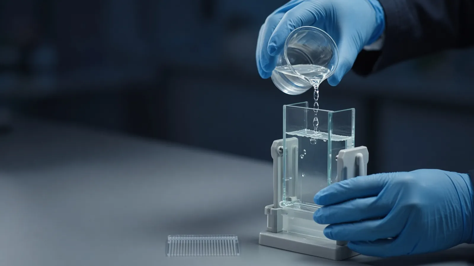
Reading a protein gel is the skill of saying what a lane can support and stopping there. SDS-PAGE, the common denaturing gel, separates proteins largely by polypeptide length. A band at the expected position is a clue. It is not a name, a purity certificate, or a concentration. This page is the pillar for that reading. How dark the band is, and how laboratories estimate protein concentration in solution, is the companion guide how laboratories estimate protein concentration. The page is a research explainer, not a clinical electrophoresis protocol.
Who uses the gel, and for which decision
A gel answers practical questions. Did the expression culture produce a polypeptide near the mass you designed? Did the purification step remove the neighbours? Did the sample enter the gel at all? Structural biologists, assay developers and cloning labs all ask versions of those questions. The decision after the gel is whether to pool a fraction, repeat a lysis, or send the band for a more specific test.
The sequence mass comes from the construct you actually expressed, including tags and cleavage scars. UniProt is a public place to check the unmodified mass of a named protein and to see known processing. Your plasmid map still wins if you added a tag the database entry does not have.
Why the proteins move
In a typical SDS-PAGE sample buffer, sodium dodecyl sulfate binds along the chain and overwhelms most of the protein's own charge. A reducing agent such as a dithiothreitol or beta-mercaptoethanol class reagent breaks disulfide links so subunits and knotted domains can unfold. Heat finishes the denaturation. In an electric field the coated polypeptides move toward the anode, and the polyacrylamide mesh sieves them so longer chains lag behind.
That story fails in informative ways. Membrane proteins and very acidic or basic proteins can bind SDS unevenly. Incomplete reduction leaves a disulfide-linked species that runs elsewhere. A native gel, run without SDS, separates by size and shape and charge together. Do not read a native lane with SDS-PAGE habits.
The acrylamide percentage sets the window. A higher percentage resolves smaller proteins and can exclude large ones from the resolving gel. A lower percentage does the opposite. Gradient gels span a wider window. The stack, the buffer system, and the voltage are part of the method. A smeared lane is sometimes a gel that was run too hot, and sometimes a sample that was never a single species.
What you load, and what you stain
The ladder is a set of proteins of stated masses. Unstained markers used with a total-protein stain are the fairer size comparison. Prestained markers are convenient and can migrate differently from the masses printed on the card. Record which ladder you used.
How much protein you load depends on a quantification you trust. A Bradford assay is one common dye-binding estimate, and it is not interchangeable with a band's darkness. Load a known mass when you can. Loading "20 microlitres of each fraction" compares volumes, not protein, unless the concentrations match.
Stains differ in sensitivity and in how honestly they report amount. Coomassie-type dyes are the everyday choice and stay more linear over a useful range. Silver stains see less protein and are easier to saturate, so a dark silver band is a poor ruler. Fluorescent stains have their own linear window and need an imager, not a phone photograph taken at an angle. Whatever you use, the image must show the whole lane, the ladder, and the dye front.
Equipment classes are ordinary: glass plates or a precast cassette, a comb, a tank, a power supply, and a staining tray. Precast and hand-cast gels can both work. They are not the same percentage just because the box looks similar. Public method notes on protocols.io and cloning-lab orientations such as Addgene's protocol collection show how laboratories document a gel. Follow the safety sheet for the acrylamide you actually pour. Vendor catalogues, including the NEB product pages, illustrate reagent classes. They are not a recipe for your tank.
A lane-by-lane reading
Start with the ladder. If the marker is curved, compressed, or missing at one end, the run itself is suspect and every sample lane inherits that doubt. Then look at whether colour from the sample even entered the resolving gel. Protein trapped in the well can be aggregated, insufficiently denatured, or simply too large for that percentage.
A sharp band near the expected mass supports "a species of about this size is abundant". Extra bands support "other polypeptides are present". They do not tell you whether those extras are breakdown products, subunits, or unrelated proteins from the host. A smear from the well to the dye front often means degradation or a grossly overloaded lane. Repeat with less protein before you invent a new isoform.
Compare lanes only when the loads are comparable and the image is not saturated. A darker band can mean more protein, a protein that stains better, or a lane that was overloaded into a non-linear stain. Quantification on a gel needs standards of the same protein, or at least a stated assumption, and a stain that has not hit its ceiling.
| What you see | A careful reading | What it does not prove |
|---|---|---|
| One sharp band near the expected mass | The sample is enriched for a polypeptide of about that size | Sequence identity or biological activity |
| Several sharp bands | More than one species is abundant | Which band is the construct |
| Material stuck in the well | Some protein did not enter this gel | That the protein is absent |
| Empty lane | Little stainable protein was loaded, or it ran off | That the culture failed, until loading is checked |
| Smiling or warped marker | The run was uneven | A new molecular weight for the sample |
Failure modes
Overloading is the most common self-inflicted mystery. The band blooms, neighbours disappear into the smear, and the gel looks "dirty" when it is merely too full. Underloading makes a successful purification look empty. Both are reasons to revisit protein quantification before the next gel.
Incomplete heating or a reducing agent that has oxidised in an old bottle leaves disulfides intact. A band that appears only in the non-reduced lane is a useful clue, not a broken gel. Keratin from skin and dust is a classic extra band on sensitive stains. Gloves and covered tubes matter more as the stain gets more sensitive.
If the dye front runs off the gel, small proteins may have left with it. Stop the run while the front is still on the gel when those masses matter. If the power supply restarts after a cut, the bands can be broader than a continuous run. Note the interruption on the image record.
Safety
Unpolymerised acrylamide is a hazardous monomer. Weighing and pouring it belong under the chemical controls your institution sets, with the gloves and waste path those rules require. Polymerised gels are handled as chemical waste, not as ordinary trash. Reducing agents smell and irritate. This article does not authorise a diagnostic gel or a biosafety decision about the source organism. The culture's containment is a separate institutional call.
Heat, humidity and shared tanks
In a hot room, a gel run at the same displayed voltage can warm enough to distort bands. Lower the current or cool the tank if the plates are too hot to touch comfortably, and record what you changed. Humidity curls dried gels and fogs imagers. Dry the gel the way the stain protocol states, and store the image with the notebook, not only on a camera roll. Precast gels follow their own expiry. A cassette that has dried at the edges will smile even when the sample is perfect.
What to send with an enquiry
Name the protein mass you expect, the expression host, whether you need a hand-cast or precast format, the stain class, and whether the gel is a purity check or a step before a blot or mass spectrometry. The reagents and chemicals catalogue is the place to start a sourcing list. The custom protein expression reference is an independent method page if the gel is part of a larger expression question. Ask through the quote request whether a quotation is possible. Do not read that page as a statement that EVRINTH runs the expression or holds a particular gel kit. A catalogue family name is not a percentage and not a stain recipe.
Read an SDS-PAGE gel without over-claiming
- 01Write the expected mass and the gel percentageLook up the sequence mass, note whether the sample was reduced, and record the acrylamide percentage. A band can only be compared with that plan.
- 02Load a ladder and a known amountInclude a marker that brackets the expected size and a load you can relate to a quantification method. Equal volumes of unknown concentration are not equal protein.
- 03Describe the lane before you name the proteinRecord the dominant band, extra bands, material in the well, and the dye front. Identity needs an orthogonal check such as a blot, activity, or mass spectrometry.
- 04Decide the next measurementIf the lane is empty, revisit extraction and loading. If many bands are present, the gel has done its job by showing impurity. Quantification of a mixture is a separate assay.
Questions from the bench
Does a band at the expected size prove the protein is pure and correct?
No. Many proteins share a similar mass. A single band is consistent with enrichment and still compatible with a co-migrating contaminant. Identity needs a specific antibody, activity, or mass-spectrometric evidence.
Why did my protein run higher than the sequence mass?
Glycosylation, other modifications, unusual amino-acid composition, incomplete denaturation, or a tag can change migration. Prestained markers are also approximate. Treat the ladder as a guide and check the chemistry of the sample.
Can I judge concentration from how dark a Coomassie band is?
Only inside a careful standard curve on the same gel, and only for a stain that is still in its linear range. A Bradford assay or another solution assay answers a different question and is explained in the companion article on protein concentration.
Is a research protein gel a clinical test?
Not by itself. A gel in a research notebook supports a biochemical claim about that sample. Diagnostic protein electrophoresis has its own validation and reporting rules.
References
Manufacturer names identify published method classes. Trademarks remain with their owners. Catalogue records on this site are independent references for enquiry. They are not a statement of inventory, distribution rights or a supply commitment. This page is educational. It is not medical advice, a diagnostic protocol or a biosafety approval.
Catalogue
Related products and categories
These links follow the subject of the article into published manufacturer references. A listing is a reference for an enquiry, not a statement of stock or distribution rights.
Continue in this cluster
Related reading
 How laboratories estimate protein concentrationHow laboratories estimate protein concentration with absorbance, dye-binding and copper assays, and why the number depends on the standard.A glossary of protein-prep termsFourteen protein-prep words, from lysate to glycerol stock, each tied to the mistake it stops before a purification is misread.Activity assays versus abundanceMilligrams per millilitre are not enzyme units. Specific activity joins rate and mass, and a pure inactive protein differs from a crude active one.