
protocol overview
Detergents salts and why they matter
Why SDS, other detergents and leftover salts suppress electrospray, which cleanup classes remove them, and how a flat ion current with a normal UV trace should
- Author
- EVRINTH Editorial Team
- Published
- 8 October 2026
- Updated
- 8 October 2026
- Reading time
- 10 min
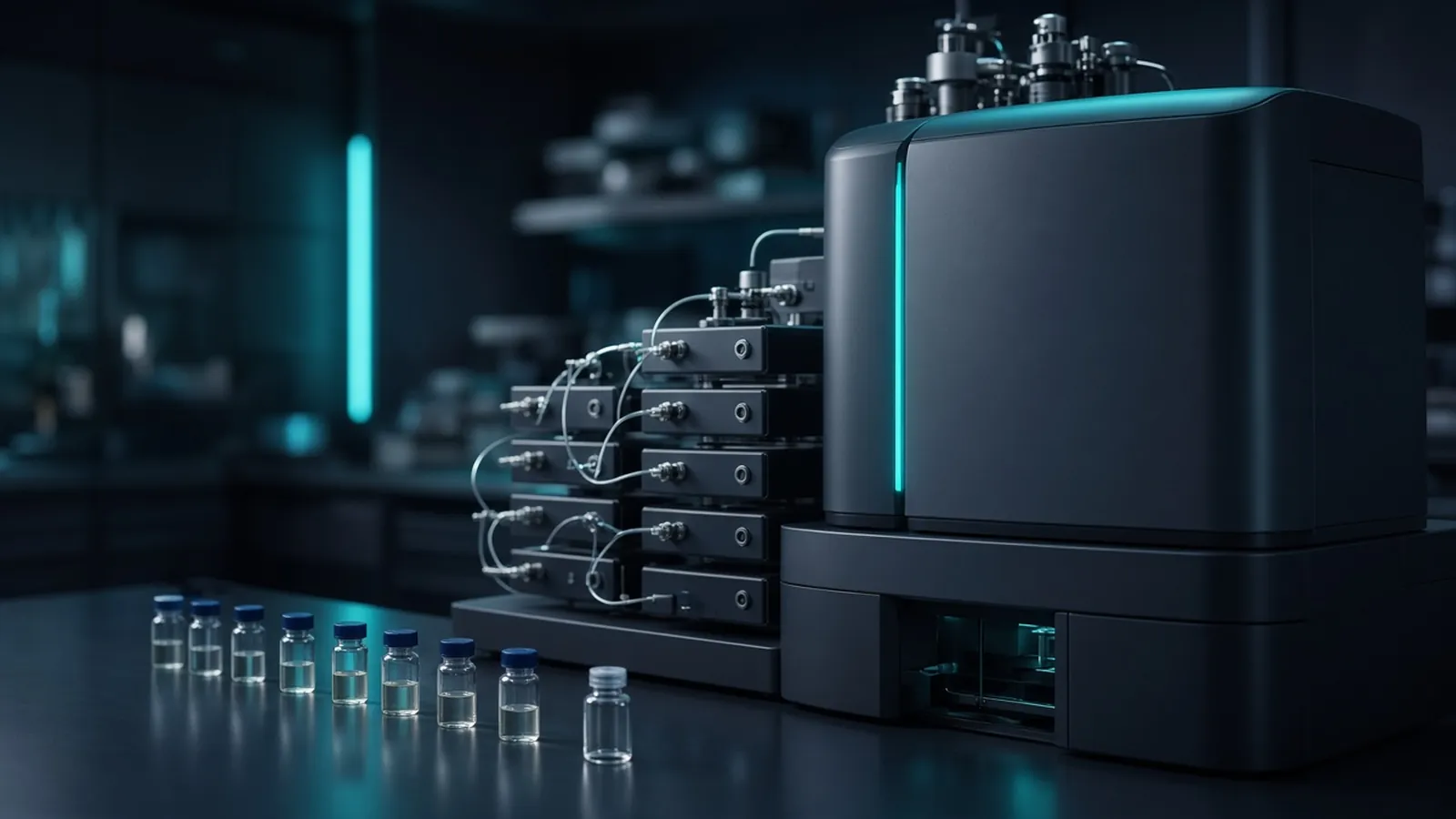
Detergents and salts decide whether a careful digest ever becomes a spectrum. They ride with peptides into the electrospray, take charge for themselves, and broaden or kill the reversed-phase separation. Bottom-up proteomics in plain language explains how identifications are claimed once ions exist. This page is the earlier decision: which additives your lysis buffer is allowed to contain if the peptides must be sprayed.
Ionisation is a competition for charge
Electrospray asks the liquid at the emitter to become charged droplets, then gas-phase ions. Peptides do this well when the droplet is mostly solvent, a little acid, and peptide. Sodium, potassium, phosphate and the hydrophobic tails of detergents also want that charge. When they win, peptide signal drops even though the vial is full of analyte. The pattern on the spectrum is often adducts, a polymer series, or a high chemical background with few assigned peaks.
Sodium dodecyl sulfate is the harsh case. It solubilises membranes brilliantly and then refuses to leave. Concentrations that are still dilute by everyday biochemistry, often discussed from about a hundredth of a percent upward, are already enough to damage a reversed-phase peptide separation and the spray. Treat any visible SDS as something that must be removed by a method written for SDS. Do not discover the limit by injecting a lysis aliquot "just to see".
Non-ionic detergents such as Triton-type and NP-40-type surfactants are polyethylene-glycol relatives. They produce a ladder of peaks separated by the repeating unit of the polymer. That ladder is a fingerprint. It also suppresses peptides across a wide stretch of the chromatogram. A buffer recipe copied from a western blot or an immunoprecipitation kit often contains one of these. The kit was solving a different problem.
Surfactant classes that can be compatible, if the removal is real
Some workflows use an acid-labile surfactant. The molecule helps the digest, then low pH breaks it, and the pieces are removed or cease to act as detergent. The class is compatible only when that acid step is performed as the protocol describes. Naming the class on a slide does not remove it.
Sodium deoxycholate is a different bargain. It solubilises well, and many proteomics protocols precipitate it with acid, often a strong drop in pH, so the detergent falls out and peptides stay in solution. The precipitation has to be complete, and the supernatant has to be taken without the pellet. A half-cleared tube is still a detergent sample.
Chaotropes are not detergents, and they cause the same class of trouble if they reach the column. Urea at several molar, and guanidinium salts at the high molarities used to dissolve stubborn proteins, must be diluted before trypsin is happy and removed before reversed-phase chromatography. Guanidinium is particularly unkind to trypsin, so dilution or buffer exchange before the enzyme is part of many protocols. Urea that sits warm carbamylates lysines. Keep those steps cold and short, and follow the manufacturer's times rather than a remembered overnight.
Water quality sits underneath all of this. A buffer made in poor water brings its own ions. Laboratory water types and where they fail is the upstream check when blanks already look salty.
Cleanup families, and the peptides they can lose
Precipitation with organic solvent or acid crashes proteins, and you wash the pellet. It can remove SDS and salts. It can also lose small proteins and leave hydrophobic proteins stuck to the tube. A pellet that was washed too hard is a changed proteome.
Bead-binding methods in the style of SP3 keep proteins or peptides on a paramagnetic surface while detergents and salts are washed away, then release the material for digestion or injection. They are attractive for small amounts. The wash solvents and the binding conditions are the method. A bead protocol copied halfway will either fail to bind or fail to release.
Suspension-trap methods, often called S-traps in the literature, combine a particulate trap with a wash that strips SDS, then digest proteins held in the trap. They are a genuine SDS-removal class. They are also a specific device and a specific solvent sequence. Follow that sequence.
C18 desalting is the last common step, and it is the wrong tool to ask for miracles. A C18 tip or cartridge retains peptides in aqueous acid and elutes them in high organic solvent, leaving many salts behind. It is the right end step after a digest that is already free of stubborn detergent. It is a weak plan for a tube that still contains SDS. Preparing peptides for mass spectrometry walks the desalting logic in more detail.
Every cleanup that removes detergent can also remove hydrophobic peptides. If your biological question lives in membrane proteins, look at which peptides disappeared, not only at whether the spray recovered. A beautiful total-ion current of soluble peptides can be a biased sample.
When the ultraviolet trace and the ions disagree
Run a solvent blank, then a control digest you trust, then the real sample. Watch two pictures. Ultraviolet absorbance, often near the peptide-bond wavelengths a method specifies, reports material that absorbs light. The total-ion current reports material that became ions.
If both are flat, the peptides may never have been there: a lost pellet, a bead elution that failed, a digest that was never loaded. If absorbance looks like a peptide run and the ion current is flat, suspect suppression or a column that is still contaminated. A detergent-poisoned column can make every later sample look empty, including a standard that worked last week. That is the branch: take the column out of service or wash it by the maker's guidance, and confirm with the standard before another real injection.
If the ion current is strong but the search finds detergent polymers and little else, the cleanup did not remove the surfactant. Repeating the same C18 step is unlikely to be the fix. Go back to a removal class matched to the detergent.
Contaminant, symptom, and the removal class that fits
| Contaminant | What you tend to see | Removal class that is actually aimed at it | A cleanup that often disappoints |
|---|---|---|---|
| SDS | Failed spray, ruined reversed-phase peaks, later injections also sick | Precipitation, SP3-style beads, suspension trap | A C18 tip used as the only step |
| PEG-type non-ionic detergent | Repeating polymer series, broad suppression | Methods written to drop that detergent before digestion, or beads with a proven wash | Dilution alone |
| Deoxycholate | Suppression until the acid precipitation is complete | Acid precipitation as the protocol describes, then desalting | Skipping the acid step because the digest "looked clear" |
| Urea or guanidinium salt | Adducts, poor retention, trypsin that also stopped working | Dilution before the enzyme where required, then reversed-phase desalting | Injecting the digest neat |
| Buffer salts | Sodium or potassium adducts, unstable spray | C18 or equivalent desalting into acidified water and organic eluent | A faster spin with no wash |
PRIDE archives show how often methods sections name these cleanup classes beside deposited runs. Read them as examples of reporting, and still follow the protocol in your hand. NIST material on measurement quality is a reminder that a suppressed signal is a measurement failure, not a small biological difference.
Failure modes worth separating
Suppression and an empty tube look alike if you only stare at the ion current. The ultraviolet trace, a peptide assay after cleanup, and a spiked control peptide are the ways to separate them. Promega protocols include protein and peptide handling notes of this general kind. Use them as method-class reading. Match volumes to the product you have.
A column that was hit with SDS can take a long time to stop bleeding. If a blank after a detergent accident still shows the polymer or still kills a standard, the column is the sample. Replacing or thoroughly cleaning it is cheaper than reinterpreting ten empty files.
Hydrophobic peptide loss shows up as a systematic absence of membrane proteins, or of the late-eluting peptides you used to see, after a precipitation that otherwise "worked". Soften the wash, change the cleanup class, or compare with a method known to keep that protein class. Do not average the loss into a pathway story.
Residual acid and residual organic solvent after cleanup change the injection. A tube eluted in strong organic solvent and injected onto a trap in a large volume may not bind. Dry or dilute into the loading solvent the chromatography method expects. That failure is physical, and it masquerades as suppression.
Solvents, acids, and institutional limits
Acetonitrile, methanol, trifluoroacetic acid and formic acid are the everyday hazards of peptide cleanup. They are flammable or corrosive as the safety data sheet says. Work in the ventilation your institution specifies, and keep concentrated acid out of the mass spectrometer's solvent bottles at the wrong strength. A research cleanup is sample chemistry. It is not a clinical preparation, and it does not authorise injection of material from an infectious sample just because the proteins were precipitated. Containment is your biosafety committee's decision.
Humidity, and a pellet that only looks dry
In humid air a centrifugal evaporator slows down, and a pellet that looks dull can still hold a film of salt and solvent. That film suppresses the spray the next morning and can leave a crystalline ring you only notice when the tube refuses to dissolve cleanly. Dry to the endpoint the protocol describes, check that the tube is truly free of liquid, and dissolve in the loading solvent while the peptides are fresh. Do not stopper a half-dry tube and leave it because the evaporator queue is long. If the room is hot as well as humid, solvent left in the tube keeps reacting with residual acid. Freeze a finished, dry sample or inject it. protocols.io records many drying steps; the climate those steps assume may be cooler and drier than the room you are standing in.
What an enquiry should say about the lysis buffer
Name the detergents already in the lysis buffer, not only the biological source. SDS, a PEG-type detergent, deoxycholate, urea and high salt each force a different removal class. State the protein amount class, whether the proteins are membrane-rich, and whether a later enrichment such as phosphopeptides must survive the cleanup. Ask which surfactant class, if any, is compatible with the mass spectrometry method you need, and which removal step is obligatory.
Those choices can be discussed with the shotgun discovery proteomics reference, the protein identification by LC-MS/MS reference and the differential abundance reference. Put the lysis recipe in the quote request. A method discussion starts from the additives you already have. It does not start from a hope that any detergent will be invisible to the spray.
Decide how a lysis additive will leave the sample
- 01Write every surfactant and salt already in the tubeList SDS, polyethylene-glycol detergents, deoxycholate, chaotropes and the buffer salts by class. A cleanup can only be chosen against that list.
- 02Pick a removal class that matches the additiveUse precipitation, bead binding, a suspension trap or an acid-cleavable surfactant workflow only if the protocol includes the removal step. A C18 tip removes many salts and does not reliably clear SDS.
- 03Compare a UV trace with the ion currentIf ultraviolet absorbance looks like peptides and the total-ion current is flat, treat suppression or a contaminated column as the leading explanation. Inject a solvent blank before you blame the digest.
- 04Retire or wash a column that has already seen detergentA reversed-phase column that received SDS can keep bleeding surfactant into later injections. Wash by the column maker's solvent guidance or take that column out of the peptide queue.
Questions from the bench
Can a C18 desalting tip remove SDS?
Do not plan on it. Reversed-phase tips are excellent at parting peptides from many salts, and SDS sticks and co-elutes in ways that still ruin the spray and the next chromatogram. SDS wants a removal class written for it, such as precipitation, a bead method or a trap chemistry the protocol validates. Follow that protocol rather than adding extra washes until the tip runs dry.
Are acid-labile surfactants safe for mass spectrometry by themselves?
The class is designed so that acid breaks the surfactant into pieces that can be removed or that no longer behave like intact detergent. The removal or degradation step is part of the method. Skipping it leaves you with the same suppression you were trying to avoid. The supplier protocol is the authority for acid strength and time.
Why did the UV chromatogram look fine while the mass spectrometer was silent?
Ultraviolet absorbance at peptide wavelengths reports chemical bonds, not gas-phase ions. Detergent, salt or a failing spray can leave the absorbance trace looking populated while few peptides ionise. A column still coated with surfactant produces the same split. Read the two detectors together before you repeat the digest.
What should a lysis-buffer enquiry say?
Name the detergents already required for solubilisation, the salt and chaotrope classes, the protein amount class, and whether membrane proteins must stay soluble until digestion. Ask which removal step belongs with that recipe. The conversation can cover compatible reagent classes. It should state the additives in the tube, not a catalogue family name alone.
References
Manufacturer names identify published method classes. Trademarks remain with their owners. Catalogue records on this site are independent references for enquiry. They are not a statement of inventory, distribution rights or a supply commitment. This page is educational. It is not medical advice, a diagnostic protocol or a biosafety approval.
Catalogue
Related products and categories
These links follow the subject of the article into published manufacturer references. A listing is a reference for an enquiry, not a statement of stock or distribution rights.
Continue in this cluster
Related reading
Bottom-up proteomics in plain languageBottom-up proteomics in plain language: proteins are digested to peptides, a mass spectrometer fragments them, and a database search names candidates. Laboratory water types and where they failResistivity, organic carbon and endotoxin answer different questions. A water grade fails when the assay cares about a contaminant the meter cannot see.Missed cleavages and ragged endsHow an internal lysine or arginine differs from a ragged peptide end, what a high missed-cleavage rate says about the digest, and how search settings affect the