
protocol overview
Enzymes lose activity before they look degraded
An enzyme can lose activity while the Coomassie band remains. Oxidation, carryover, a missing cofactor or partial unfolding need an activity assay.
- Author
- EVRINTH Editorial Team
- Published
- 8 October 2026
- Updated
- 8 October 2026
- Reading time
- 7 min
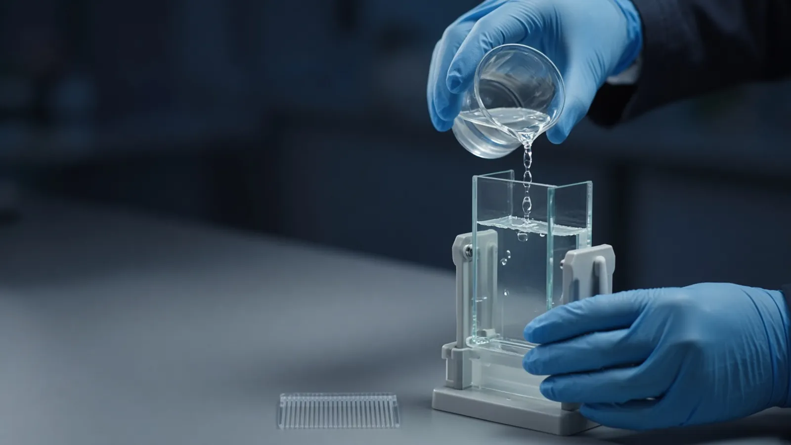
Catalysis can fail while the polypeptide chain is still the length the gel expects. A Coomassie band that looks as strong as last month's does not carry last month's specific activity with it. Active-site oxidation, inhibitor left over from the purification, a cofactor you forgot to add, and a partial unfold are common ways an enzyme goes quiet without looking degraded. This overview is the decision path for that disagreement. How mass and rate share a sentence is continued in activity assays versus abundance. Inhibitor carryover is named in protease inhibitors and when they interfere. The stained lane itself is interpreted in reading a protein gel.
The band is abundance of stain, not a rate
SDS-PAGE with a total-protein stain shows chains that are present and soluble in sample buffer. It is almost silent about the geometry of an active site. A nick that removes three residues from a terminus may not move the band enough to notice, and a chemical change to one cysteine may not move it at all. People still write "the protein looks fine" under a photo and shelve the assay. The photo answered a mass question. The experiment asked a catalytic one.
Specific activity puts them together: product formed per time, divided by the mass of enzyme, under stated conditions. If the mass is an albumin-equivalent Bradford number of a mixture, say so. If the mass is from a coefficient, the coefficient's assumptions apply. Either way, a drop in specific activity with an unchanged band is a real result. It means something in the catalytic machinery, the assay, or the soluble fraction changed.
Four mechanisms that spare the gel
Oxidation is the quiet one. Catalytic cysteines, iron-sulfur chemistry and some other centres lose function when they oxidise. The backbone is intact. A freshly prepared enzyme and one that sat in a thin film of liquid overnight can share a gel and not share a rate. Sometimes a reductant restores the cysteine enzyme. Sometimes the damage is past that. Add reductant only if the enzyme's instructions allow it, because a reductant is also an assay interference for many copper methods and for some substrates.
Inhibitor carryover is the rude one. PMSF or AEBSF left in a serine protease, leupeptin left in a cysteine protease, or EDTA left in a zinc enzyme will suppress activity. The gel remains flattering. Buffer exchange, done until conductivity and the assay agree, is the test. A spike of uninhibited control enzyme into the same buffer shows whether the buffer itself is poisonous.
Missing cofactor is the embarrassing one. Kinases need the right nucleotide. Many hydrolases and oxidoreductases need a metal or a prosthetic group that purification removed. UniProt often lists cofactors for a characterised enzyme. Add back only what that enzyme is known to require, at a range from its protocol, and include a no-enzyme blank so the cofactor is not mistaken for activity. Do not dump a random metal mix into the well.
Partial unfolding is the structural one. The chain is complete, the hydrodynamic behaviour may already be wrong, and the active site no longer forms. A cloudy sample, a void peak, or a protein that has been through too many freeze-thaws belongs in this bin. Clarifying and assaying the supernatant tells you whether any folded material remains. A band in the well after a native handling, compared with a normal SDS band, supports the picture. It still does not replace the rate measurement.
An assay at decision level
Define the product. If the reaction releases a coloured or fluorescent species, build a standard curve of that product in the same buffer. Read unknowns inside the curve. A blank without enzyme catches substrate breakdown and instrument drift. A blank without substrate catches enzyme-only colour. Time points, or a continuous rate in the linear window, beat a single endpoint that may already have plateaued.
Temperature and pH are part of the unit. An assay run at an unspecified "room temperature" in a hot laboratory is not comparable to one run at a controlled 25 or 37 Celsius. Use the range the enzyme's instructions give, and write the number you held. Substrate concentration relative to the enzyme's typical working range matters. Too little substrate makes every enzyme look slow. This article does not give a microlitre recipe. Follow the source protocol for volumes, and keep the logic of blank, standard and linear window.
If the first assay is zero, branch. Confirm the gel still shows intact protein in that aliquot. Confirm the blank is low. Then test carryover, cofactor and a fresh dilution. A zero from an assay with a high blank is an uninterpretable assay, not a dead enzyme. A zero from a clear assay, an intact band, no inhibitor and the right cofactor is when you earn the right to discuss unfolding or a failed refold.
| What you observe | Gel | Assay | The next question |
|---|---|---|---|
| Intact band, high blank | Chain is there | Uninterpretable | Fix spontaneous signal before judging the enzyme |
| Intact band, true zero | Chain is there | No product | Inhibitor, cofactor, oxidation, or unfold |
| Intact band, lower rate | Chain is there | Partial loss | Storage history, partial oxidation, mix of inactive protein |
| Fragments, low rate | Chain is cut | Likely real loss | Proteolysis, earlier inhibitors, time and temperature |
| Cloudy tube, odd rate | May be in the well | Mass may be wrong | Clarify, then recalculate per soluble milligram |
Failure modes that masquerade as biochemistry
An expired substrate looks like a dead enzyme. Test the substrate with a trusted aliquot if you have one. A coupling enzyme that has itself died looks like the primary enzyme died. Check the coupling step alone. Evaporation in a warm plate raises concentrations mid-assay and bends the curve. Seal the plate and control the temperature. Calculating a rate from a single saturated endpoint makes two different enzymes look identical. Stay in the linear window.
Densitometry of the band cannot rescue a missing standard curve. Neither can a more sensitive stain. Sensitivity is not specificity of the reaction.
Research limits, climate and enquiry
Enzyme assays in this sense are research measurements. They are not clinical enzyme tests and not a diagnostic approval. Hazardous substrates and coupling enzymes follow their own safety notes. Biosafety of the source material is institutional.
Ice buckets in a hot room are a short story. An assay that says "on ice" and then waits an hour in meltwater has left the condition you think you held. Power cuts stop plate readers mid-kinetic. Discard that curve and rerun it. Record the room and the instrument temperature when the rate is temperature-sensitive. A frost-free freezer that cycled all week can be the entire explanation of a lower specific activity. Check storage history before you redesign the enzyme.
An enquiry about assay reagents should name the enzyme class, the product you will detect, any cofactor, and the inhibitor classes the sample may still contain. Components sit in the reagents and chemicals catalogue. Use a quote for that specification. If the inactive preparation is part of a broader expression question, the custom protein expression and purification page is an enquiry reference where the method can be discussed. It is not a claim that an enzyme is being produced or assayed as a service.
Separate a dead active site from a missing band
- 01Run the gel and the assay on the same aliquotStain a defined load and measure activity from the same tube on the same day. A band from last week does not vouch for a rate measured after a thaw.
- 02Include a blank and a product standard curveThe blank contains everything except the enzyme, or everything except the substrate, as the assay logic requires. Convert signal into product with a standard curve of that product. A raw absorbance is not a rate.
- 03Check carryover, cofactor and oxidation before you blame the foldAsk whether an inhibitor, a chelator, a missing metal or nucleotide, or an oxidised cysteine could silence a still-intact chain. Remove the suspect or add back the cofactor the enzyme is known to need, and repeat.
- 04Report specific activity with the conditionsWrite the rate per mass, the temperature, the pH and the substrate definition you used. Follow the enzyme's own instructions for ranges. Do not paste an unrelated kit recipe into the notebook.
Questions from the bench
Can a Coomassie band prove an enzyme is active?
No. The stain reports polypeptide that entered the gel. Active-site oxidation, a bound inhibitor, a missing cofactor or a partial unfold can remove activity with little change in mass. Specific activity is a different measurement, explained alongside abundance in the companion article.
What does a blank actually subtract?
It subtracts signal that appears without the reaction you claim. Substrate that breaks down on its own, a coloured buffer, or a coupling enzyme that drifts will otherwise look like product. If the blank is as high as the sample, fix the assay before you conclude the enzyme is absent.
Why would oxidation kill activity and leave the gel unchanged?
A cysteine in the active site can oxidise, or a metal can change oxidation state, without cleaving the backbone. The chain length stays the same, so the band stays put. A reducing treatment sometimes restores that class of damage and sometimes does not. Test it deliberately rather than adding reductant to every assay by habit.
Are published assay recipes safe to copy in microlitres?
Treat them as decision guides. Substrate concentration, pH and temperature must sit where that enzyme works, which the enzyme source or a methods paper states as ranges. Copying another enzyme's table is how you measure a zero that the protein did not earn. This overview stays at that decision level on purpose.
References
Manufacturer names identify published method classes. Trademarks remain with their owners. Catalogue records on this site are independent references for enquiry. They are not a statement of inventory, distribution rights or a supply commitment. This page is educational. It is not medical advice, a diagnostic protocol or a biosafety approval.
Catalogue
Related products and categories
These links follow the subject of the article into published manufacturer references. A listing is a reference for an enquiry, not a statement of stock or distribution rights.
Continue in this cluster
Related reading
Reading a protein gelHow to read a protein gel: what SDS-PAGE bands, smears, ladders and loading differences can support, and what they cannot identify.Activity assays versus abundanceMilligrams per millilitre are not enzyme units. Specific activity joins rate and mass, and a pure inactive protein differs from a crude active one.Protease inhibitors and when they interfereProtease inhibitor classes stop different enzymes. EDTA, PMSF, AEBSF, pepstatin and leupeptin can also block the activity or the resin you wanted.