
guide
Fluorescence microscopy without the myths
How a fluorescence microscope selects excitation and emission, and which bright pictures still fail as evidence of a labelled molecule.
- Author
- EVRINTH Editorial Team
- Published
- 8 October 2026
- Updated
- 8 October 2026
- Reading time
- 10 min
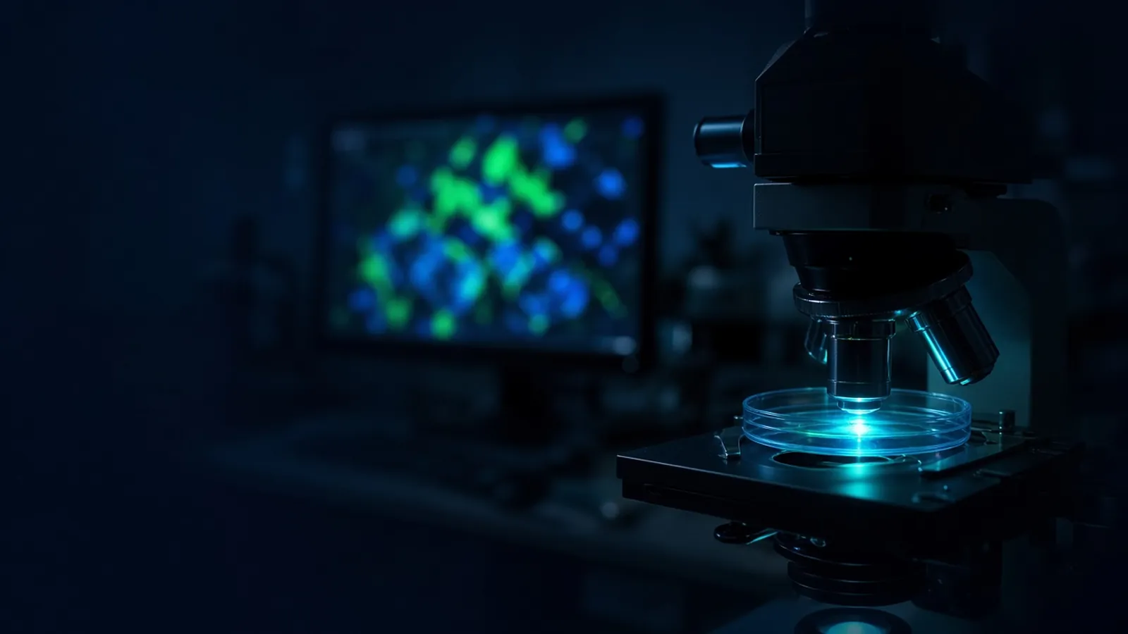
Fluorescence microscopy without the myths means treating a glowing picture as an optical measurement with controls, not as a photograph that speaks for itself. The decision this page supports is whether a bright cell, a colocalised pair of colours, or a "negative" field can carry the biological sentence you want to write. Counting the same kind of label across thousands of cells, without an image, is the job of a cytometer, explained in how a flow cytometer reads a cell. This page stays with the microscope.
Instrument classes live in the scientific instruments catalogue. The experiment often belongs to the molecular biology pathway. A specification is a quote request, not an assumption that a listed family is configured for your dyes.
What the image is allowed to decide
A fluorescence micrograph can show that, under a stated excitation and emission window, some structures emitted more light than a proper negative control. It can support a comparison of localisation when the resolution and the sampling match the claim. It cannot, by itself, prove that an antibody bound only the intended protein, that two proteins touch, or that the brightness is a concentration. Those sentences need extra evidence: a blot, a genetic control, a spectrum, or a calibrated standard. Write the sentence first. Then ask whether the optic can reach it.
Excitation, emission and the cube
A fluorophore absorbs photons in one band and, after losing a little energy, emits photons at a longer wavelength. That gap is the Stokes shift. The microscope uses it. An excitation filter or a laser line selects the light that should be absorbed. A dichroic mirror reflects that excitation toward the objective and passes the longer emission back. An emission filter blocks leftover excitation and passes the band you chose to collect. The objective both illuminates the sample and gathers the emission. Its numerical aperture, more than the magnification printed on the barrel, sets how much light you collect and how fine a detail you can hope to separate.
Widefield illumination lights a thickness of sample. Light from above and below the plane of focus still reaches the camera, which is why a thick tissue slice looks foggy. A confocal instrument rejects much of that out-of-focus light with a pinhole. It does not invent resolution the objective does not have, and it does not remove autofluorescence that is truly in the focal plane. "Confocal" is a way of rejecting blur, not a certificate of specificity.
The camera or detector integrates photons for an exposure, or it counts them. Gain multiplies signal and noise together. Saturation is a hard failure: pixels at the maximum value have no intensity left to compare. A figure in which the interesting structure is a flat white shape cannot support a claim about "more" or "less".
Myths that change the conclusion
The brightest image is the truest image. Brightness is photons times gain times exposure. You can make a negative sample look positive by lingering. The honest comparison uses identical excitation power, exposure and gain, and shows the negative next to the positive. If you must adjust, say so, and do not hide the adjustment in a contrast tool after export.
A colour is a molecule. Green is a filter, not a substance. Enhanced green fluorescent protein, fluorescein-like dyes, and cellular autofluorescence can all occupy that window. Red is no safer: chlorophyll, fixative artefacts and long-Stokes dyes collide there too. Fluorescent proteins themselves are reagents with maturation times and a tendency to bleach. Cloning notes gathered by Addgene are a place to see how those proteins are handled as genetic tools. They do not make a particular cell glow on command.
Overlap of two colours means the molecules interact. It does not. It means both emissions were collected from volumes the microscope could not tell apart. In a widefield image of a thick cell, that volume is large. Even in a confocal image, diffraction limits how close "together" is. Interaction is a biochemical claim. The image can be consistent with it and still be wrong.
The eye and the camera saw the same thing. The camera integrates, false-colours, and displays a lookup table. A lookup table that paints dim grey as bright gold changes the rhetoric of the figure without changing the file. Keep the raw values. State the display bounds.
Fixation freezes the biology exactly as it was. Cross-linking and permeabilisation move soluble proteins, kill some epitopes, and create autofluorescence. A live-cell image and a fixed image of the same reporter can disagree for optical reasons as well as biological ones. Do not mix them in one claim.
Compensation, in the cytometry sense, has been done because the software has a slider. Microscope software often has brightness and a bit of spectral unmixing on newer systems. Unmixing needs reference spectra taken on that instrument. A slider is a display choice. The cytometry companion page explains why overlap is a matrix, not a feeling. The same spectra are the reason a double-labelled slide needs single-labelled controls.
Equipment classes, not a brand ritual
Light sources are lamps, light-emitting diodes or lasers, chosen because their lines hit the excitation peak. Filter cubes are matched sets. Mixing an excitation filter from one dye with an emission filter from another is how you either see nothing or see everything. Objectives are dry or immersion; an oil objective used dry, or oil left on a dry objective, wrecks the image and can wreck the lens. Culture dishes and coverslips have thicknesses the objective was designed around. A plastic dish under an objective corrected for a glass coverslip is a soft, false image. Vessel choice is part of the optic.
Antibodies, fluorescent proteins and chemical stains are the labelling classes. A primary antibody without a no-primary control, on a sample that was fixed with an aldehyde, is a common source of confident wrong figures. Record the label, the dilution you actually used, and the fixative.
A session when the control glows
Set the negative specimen first. Find a field, set exposure so the background is low but not clipped to zero everywhere if you still need to prove the camera was on, and note the numbers. Move to the sample without changing those numbers. If the sample is saturated, reduce exposure. Do not "fix it" only in the export.
If the negative glows as brightly as the sample, stop. Causes include autofluorescence, a filter that passes excitation light, a stain that binds non-specifically, and contamination of the mounting medium. Changing the lookup table until the negative looks dark and the sample looks bright is how myths are published. Try a different emission window, a spectral check if you have one, or a different labelling chemistry. If single-stained controls appear in the other channel, you have bleed-through. Image those channels sequentially with one laser at a time, or accept that this pair of dyes cannot share that cube.
| Control | What you learn when it is dark | If it glows |
|---|---|---|
| Unlabelled cells, same settings | Autofluorescence is not filling the window | Do not call sample brightness specific |
| No primary antibody | The secondary is not sticking on its own | The secondary or the block has failed |
| Single fluorophore only | The other channel is quiet for this dye | Bleed-through; do not claim a second molecule |
| Known localisation control | The optic can put that label in the right place | A new pattern in the sample is still only a pattern |
Photobleaching is a branch of its own. If the signal fades while you focus, the exposure you finally save is the dim remnant. Focus with low light, or focus in a transmitted channel, then take the fluorescence frame. A time course of bleaching is not a biological disappearance unless an unilluminated sister field still has its signal.
Failure modes
Dim images with a correct control are often objective, immersion or alignment problems, or a dye excited by the wrong line. Bright images with a bright control are specificity problems. Soft images are coverslip thickness, refractive-index mismatch, or a dirty objective. A field that looks sharp on the camera and empty through the eyepiece can be a light-path selection left on the camera port, not a biological negative. Write down the cube name. Two cubes both called "green" can have different bandpasses.
Light, specimens and research limits
Excitation light, especially ultraviolet and strong lasers, damages eyes and skin. Interlocks and the correct filter, not a habit of squinting, are the control. The specimen has its own rules. A culture dish on a microscope stage is still the organism or the cell line your institution assessed. The CDC BMBL and the WHO Laboratory biosafety manual are references for that assessment. This article does not assign a containment level, does not diagnose a patient slide, and is not medical advice.
A written imaging note that records objective, filters, exposure and the controls is what makes the figure repeatable. The exported colour picture is the illustration.
Humidity, heat and shared microscopes
In a humid season, fungus grows on optics and in cameras. Dry the room as the facility allows, and do not leave oil on a lens overnight. Temperature cycling from aggressive air-conditioning shifts focus during a time lapse; an enclosure or a patient stage is part of the method, not a luxury, when the claim is a live-cell movie. Shared microscopes accumulate the previous user's immersion oil and the previous user's filter left in the path. Check the light path every session. A power cut mid-stack corrupts the z series. Start that stack again. Do not average a half-finished volume into a "representative" image.
What to send with an enquiry
Name the fluorophores, the format of the specimen, widefield versus optical sectioning, and any environmental control the live sample needs. Ask for filter bandpasses and light-source lines in writing. Use the scientific instruments catalogue and the quote request. A family name is not a demonstration that those filters are fitted.
Judge a fluorescence image against the controls that make it interpretable
- 01Name the molecule and the optical windowWrite the fluorophore or the fluorescent protein, the laser or lamp line that excites it, and the emission filter that is supposed to collect it. A colour name on a figure is not that record.
- 02Image the negative specimen with the same settingsAcquire unlabelled cells, and a no-primary-antibody control if you are using antibodies, at the same exposure or detector gain as the sample. If that control glows, the sample glow is not yet a result.
- 03Separate brightness from specificityTurn the excitation down or shorten the exposure until the negative control is dark, then ask whether the real sample is still above that background. Raising gain until something appears does not create a label.
- 04Keep spatial claims inside what the optic can resolveState the objective numerical aperture and whether the image is widefield or confocal. Overlap of two colours in a thick widefield slice is not proof that two molecules touch.
Questions from the bench
If the cell is green, is the green fluorescent protein there?
Not by colour alone. Cells autofluoresce, aldehydes used in fixation can add glow, and a neighbouring fluorophore can leak into the green filter. A green picture is compatible with a real fluorescent protein and compatible with those artefacts. The untransfected or unlabelled sister sample, taken at the same settings, is what lets you tell them apart.
Does a higher magnification make a truer image?
Magnification enlarges. Resolution depends on wavelength and on the numerical aperture of the objective, and it is also limited by the pixel size of the camera. An empty magnification that spreads a blur across more pixels looks detailed and resolves nothing new. Match the objective to the structure you claim to see.
Is bleed-through the same problem as compensation on a cytometer?
It is the same physics and a different correction. Emission spectra overlap, so a filter set collects some of the wrong dye. On a cytometer that overlap is subtracted with a compensation matrix, as described in the companion article on how a cytometer reads a cell. On a microscope you prevent it with filter choice, sequential excitation, and controls that show each dye alone. Sliding a brightness bar is not compensation.
What should an enquiry about a fluorescence microscope include?
The fluorophores you must excite, whether you need widefield, confocal or a stereo format, the sample vessel, and any camera or environmental-chamber requirement. Ask for the filter and light-source specification. A catalogue family name is not an optical layout and does not mean an instrument is sitting idle.
References
Manufacturer names identify published method classes. Trademarks remain with their owners. Catalogue records on this site are independent references for enquiry. They are not a statement of inventory, distribution rights or a supply commitment. This page is educational. It is not medical advice, a diagnostic protocol or a biosafety approval.
Catalogue
Related products and categories
These links follow the subject of the article into published manufacturer references. A listing is a reference for an enquiry, not a statement of stock or distribution rights.
Continue in this cluster
Related reading
 How a flow cytometer reads a cellHow a cytometer focuses cells into a laser, turns scatter and fluorescence into pulses, and why a gate is an argument rather than a fact.A glossary of optical measurementsWhich optical measurements a microscope or cytometer specification should name so a later user can repeat the light path.Biosafety of unfixed human samplesWhy unfixed human blood, tissue and cells on a cytometer or microscope stay an institutional biosafety decision, not a technique choice.