
comparison
Peptide fractionation overview
Single-shot runs versus offline peptide fractionation: depth, instrument time, sample use, concatenation and the losses that come with extra fractions.
- Author
- EVRINTH Editorial Team
- Published
- 8 October 2026
- Updated
- 8 October 2026
- Reading time
- 8 min
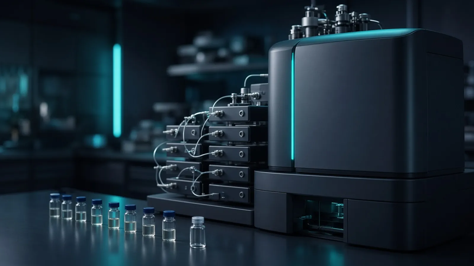
Fractionation buys a deeper look at a peptide mixture and spends three things: instrument time, sample, and the chance to lose a fraction on the way. The commercial question is which of those costs belongs in a specification for this study. It is not a ranking of brands. A simple pull-down and a whole-cell lysate do not deserve the same answer. The acquisition those fractions feed is described in bottom-up proteomics in plain language.
What an offline fraction changes
A single-shot run injects the whole digest onto reversed-phase chromatography and into the mass spectrometer once. Abundant peptides occupy the sampling. Scarce peptides are real and often unfragmented. Offline fractionation splits the digest, before that analytical gradient, into portions that are less complex. Each portion is cleaned and injected separately, or portions are recombined in a planned way and then injected. The spectrometer sees further down the abundance scale because the abundant peptides are not all in the same injection.
The split is never free. Every additional tube is a place to lose peptide on plastic, to contaminate with keratin, or to leave salt that wrecks the later gradient. Every additional injection is a place for a column to drift. A false-discovery threshold still applies to the combined search. A longer list is not automatically a truer list if the search was loosened to celebrate the extra time.
Three fraction classes
High-pH reversed phase separates peptides by hydrophobicity under basic conditions. The later analytical run is usually low-pH reversed phase. The two are similar but not identical, so a peptide's early fraction and its late analytical retention are not the same fact. This class is widely used before discovery injections because the solvents can be made compatible with mass spectrometry. The number of fractions and the gradient shape belong to the protocol you adopt. protocols.io holds many variants. Follow one protocol's loading limits rather than a blend of two papers.
Strong cation exchange separates by charge. It is orthogonal to reversed phase in a different way. Salt or pH steps elute the peptides. The salt has to come off before a nano-flow column sees the sample. A salty fraction looks like a failed analytical run: poor spray, plugs, or peptides that never bind the trap. Cleanup between the exchange step and the analytical step is part of the method, not a courtesy.
Gel slices are the crude class. A protein gel separates by approximate size. You cut bands or equal sections, digest in the gel, and extract peptides. That suits a visible band or a region you already care about. Reading a protein gel is the skill that stops you cutting the marker lane by mistake. It is a clumsy way to fractionate an entire proteome: extraction losses are large, keratin is common, and the slices are not equal peptide amounts. Use slices when the question is the band.
Concatenation, and when a single shot is enough
Concatenation is the recombination step that makes high-pH fractions practical. You might collect many fractions across the high-pH gradient, then combine fraction 1 with a late fraction, fraction 2 with another late fraction, and so on. Each tube you actually inject then contains both hydrophilic and hydrophobic peptides. The low-pH analytical gradient separates them again. You spend fewer hours on the mass spectrometer than if you injected every high-pH fraction alone, and you avoid a tube that contains only peptides that all elute in the same two minutes.
The arithmetic of how many to combine is a protocol choice. The principle is the part to specify: orthogonal split, then a stated recombination, then a stated number of analytical injections. Concatenating everything back into one tube undoes the fractionation. Concatenating neighbours rather than early-with-late puts similar peptides together and wastes the orthogonality.
A single shot is the honest specification when the mixture is already a pull-down with a modest cast of proteins, when you have many biological replicates and barely enough peptide for one good injection, or when the question is identification of a dominant band rather than a deep census. A deep proteome of a lysate, where the scientific question really is the scarce proteins, is where fractions earn their cost. "As deep as possible" is not a scientific question. "Proteins beyond the abundant metabolic set, with replicates still intact" is closer. PeptideAtlas can show how often a protein of interest has been seen at all. UniProt identifies it. Neither site will tell you that your twelve samples need eight fractions.
Studies deposited through the ProteomeXchange consortium include both single-shot and fractionated designs. Compare a deposit's fraction count with its sample count before you copy the design. A method built for three cell lines and a year of instrument time is a different specification from a comparison of two conditions.
| Choice | Depth | Instrument time | Sample consumption | Typical failure |
|---|---|---|---|---|
| Single shot | Limited by abundant peptides | One injection per sample | Lowest | The scarce protein is never fragmented |
| High-pH reversed phase, each fraction injected | Higher if loads are real | One injection per fraction | Higher, and losses on plastic | A fraction dried down and left behind |
| High-pH, concatenated | Higher than one shot, fewer runs than every fraction | A stated handful of injections | Losses plus a recombination error | Early combined with early, orthogonality lost |
| Strong cation exchange | Higher when salt is removed | One injection per fraction or pool | Peptide lost in desalting | Salt reaches the trap and the gradient collapses |
| Gel slices | Local depth in a size region | One injection per slice you keep | Extraction losses are part of the method | Keratin, empty slices, unequal loads |
A lost fraction and a salty fraction
If one concatenated tube is empty because it was left in the evaporator, the proteins that lived mostly in those fractions disappear. The rest of the list looks normal. Compare peptide amount per fraction before you inject. A tube far below its neighbours is a handling event. Recombine from remaining material if the protocol still has it, or report the missing window. Do not scale the other fractions up in a spreadsheet and call the depth complete.
A strong-cation fraction that still contains salt fails differently. The analytical chromatogram collapses or the spray is unstable, and the search from that injection is thin. Desalt again. Do not average a failed salt run with a clean one.
Uneven loads across fractions bias label-free intensity. The fraction with twice the peptide produces larger peaks for whatever it contains. Record the amounts and the injection volumes. A specification that says equal fractions needs a measurement, not a hope.
A further branch appears when the analytical column is new for the first fraction and tired for the last. Retention drifts, and a peptide quantified across fractions no longer lines up in time. Keep a short standard or the pool injection beside the fraction series so the drift is visible. If you are comparing many biological samples, prefer fewer concatenated injections per sample and more of the replicate budget. Depth that consumes the replicates answers a different question from the one the grouping was built to ask.
Search the fractions together under one false-discovery procedure when they are one sample's evidence. Searching each fraction to a loose threshold and then concatenating the protein lists inflates discoveries. The decoys have to see the same combined decision the targets see.
Research use
Fractionation is a research depth tool. It does not create a diagnostic panel and it does not assign function. Biosafety of the lysate is unchanged by how many tubes you split it into. More tubes can mean more chances to contaminate a bench. Your institution's rules still decide containment.
Many injections and an interrupted night
A fractionated queue is long. A power cut between fraction three and fraction four of the same sample splits that sample's depth experiment into two acquisition batches. Record which fractions finished, re-equilibrate, and do not describe the set as one uninterrupted measurement. In a hot, humid room, fractions waiting in the autosampler or half-dried in a concentrator pick up moisture and keratin. Cap them and keep them cold as the protocol requires. If the building power is unreliable, fractionate and inject in blocks you can finish, with the sample key written so a restarted block is labelled as a restart.
Writing depth into a specification
Write the scientific depth you need: a pull-down's partners, a gel band, or a lysate beyond the most abundant proteins. Write the peptide amount you can spend. Write whether concatenation is acceptable and how many analytical injections per sample you can justify beside the biological replicates. Ask for the failure rule when a fraction is lost.
The shotgun discovery proteomics reference is where a deep screen is discussed. The protein identification by LC-MS/MS reference fits a band or a simpler mixture. The differential abundance reference is the comparison that must still have replicates after the fractions have taken their share of the instrument. Send the depth question, not a protein-count target, with the quote request. Those pages support the discussion. They do not mean a fractionator is already assigned to your samples.
Questions from the bench
Does fractionation guarantee a longer protein list?
It often increases depth when the chemistry and the loads work, and it can shorten the list when fractions are lost or salty. Treat depth as something you measure on your mixture. Do not write a protein-count promise into a plan.
When is a single injection the better specification?
When the mixture is already simple, as in a clean pull-down, or when you have many biological samples and little peptide. Extra fractions then spend instrument time you needed for replicates.
What does concatenation change?
It combines early and late offline fractions so that each analytical run still sees a spread of peptides and you inject fewer tubes. It does not restore a fraction you dropped on the bench.
Are gel slices a fractionation method?
They are a crude separation by size before digestion. They suit a band you can see. They are a poor way to survey an entire lysate, and they invite keratin. Read the gel for what it is.
References
Manufacturer names identify published method classes. Trademarks remain with their owners. Catalogue records on this site are independent references for enquiry. They are not a statement of inventory, distribution rights or a supply commitment. This page is educational. It is not medical advice, a diagnostic protocol or a biosafety approval.
Catalogue
Related products and categories
These links follow the subject of the article into published manufacturer references. A listing is a reference for an enquiry, not a statement of stock or distribution rights.
Continue in this cluster
Related reading
Bottom-up proteomics in plain languageBottom-up proteomics in plain language: proteins are digested to peptides, a mass spectrometer fragments them, and a database search names candidates. Reading a protein gelHow to read a protein gel: what SDS-PAGE bands, smears, ladders and loading differences can support, and what they cannot identify.A glossary of proteomics termsA working glossary of proteomics terms, from precursor and PSM to razor peptide, FDR and batch effect, and how each term changes a pull-down or abundance claim.