
glossary
Photos of morphology are data
A culture photograph is a record only with passage, scale and time since seeding. This glossary separates phase contrast, confluence and morphological words.
- Author
- EVRINTH Editorial Team
- Published
- 8 October 2026
- Updated
- 8 October 2026
- Reading time
- 7 min
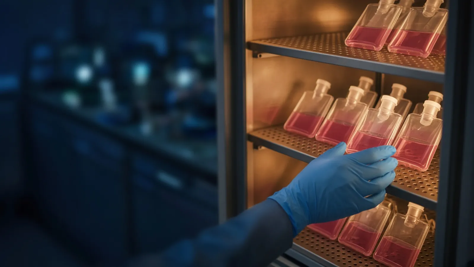
A photograph of cells is data only when a stranger can tell what they are looking at and when it was true. Without the passage number, a scale and the time since seeding, the file is an illustration. It may be a beautiful one. It cannot anchor a method. Morphology still matters: it is often the first sign that a culture has drifted, lifted or been seeded in clumps. The place of that inspection in ordinary culture work is mammalian cell culture for research labs. This page is a glossary for the picture and for the words people type under it. It is not a way to diagnose disease from a snapshot.
Optical words, before biological ones
Phase contrast is the usual method for live, unstained cultures. The microscope converts small optical-path differences into light and dark, which is why a transparent cell becomes visible. It is not a measurement of stain intensity, because there is no stain. Brightfield without phase often looks empty until the cells are dense. Fluorescence is a different channel and needs a label or a genetic reporter. Do not describe a phase image as if it were an antibody result.
A scale bar is a length drawn from a calibration, the microscope software, or a reticule you have checked. A bar you stretched until it "looked like 50 micrometres" is decoration. Magnification, if you write it, is the objective you used and any extra optovar, not a number remembered from another scope. Pixel size changes when you crop. The bar has to survive the crop.
Confluence as an image is the fraction of that field that cells appear to cover. It is an estimate. It is not cells per square centimetre unless you have calibrated the field against a count. One field is not the flask. Edges, the centre and the area near a scratch can disagree. If you report cover for the vessel, say you sampled more than one field or say you did not.
Focus artefacts are optical, and they impersonate biology. A wrong phase ring turns the monolayer into grey mud. Condensation on the lid looks like fog and hides edges. Thick plastic or a scratched flask bends the image. A halo around a rounded cell is the phase effect, not a vesicle you can count. Debris in the medium can shadow a cell and look like a granule inside it. Clean the optical path your instrument manual allows. Do not open a contaminated flask for a sharper picture.
Morphological words, used narrowly
Spread means an adherent cell has extended cytoplasm and is sitting down on the surface. It is the ordinary look of many healthy attached lines. It is not a differentiation score.
Rounded means the cell is phase-bright and ball-like. Mitotic cells round up and then spread again. Cells on the wrong plastic round up and stay there. Toxicity and a cold excursion do the same. The word rounded without the context is a shape, not a cause. Pair it with whether the cell was rounded yesterday, and with the passage and the medium.
Vacuolated means you can see cytoplasmic vacuoles, the clear intracellular bubbles, as distinct from the phase halo outside the cell. Some lines are vacuolated in ordinary culture. Some become so under stress, in an exhausted medium, or after a particular treatment. The photo shows the vacuoles. It does not name the cause. Do not write a disease name because a textbook picture of a pathology looked similar.
Lifted means cells are leaving the surface. Edges go refractile, sheets peel, or cells float as intact patches. Over-confluence, a failed coating, toxicity and rough handling all lift cells. A photo of floaters does not distinguish them. The notebook sentence next to the file has to say what changed.
Neurite-like is the dangerous compliment. A thin process can be a real neuronal projection in a protocol that used neuronal cells and the markers that protocol demands. The same silhouette on an epithelial line is a thin extension. Write neurite-like only as a shape, and only if the sentence also refuses the differentiation claim you have not earned. If you did not run the markers, do not use the word. Process is enough. Public descriptions of how a line "should" look, in the ATCC culture guides or the European Collection of Cell Cultures, are comparisons. They are not a diagnosis, and your passage may no longer match the catalogue photograph.
| What the photo shows | What it is not | Metadata it needs |
|---|---|---|
| Cover in one field | Confluence of the whole flask, or a cell count | How many fields, and the scale bar |
| A phase-contrast halo | A vacuole or a stained organelle | The method: phase, not fluorescence |
| Spread cytoplasm | Proof of differentiation or of health | Line, passage, time since seeding |
| Rounded, phase-bright cells | A named cause, mitotic or toxic | Whether they spread again, and the medium |
| Intracellular vacuoles | A disease diagnosis | The line's ordinary look at that passage |
| Lifted sheets or floaters | A specific toxin identified | What was new that day |
| A thin process | Neuronal differentiation | Markers, or the word process instead |
Taking the picture so the caption can be true
Focus on the cell layer, not on a scratch in the plastic. Use the same objective when you compare days. Record the file name in the notebook row that already holds the passage, the date and the hours or days since seeding. If you seed at a density you pipetted, the pipetting record is part of why the field looks even or clumped. Technique for that volume is a separate skill, covered in accurate micropipetting technique. A clumpy field photographed without the seed note becomes a false morphology phenotype.
Branch when the image and the eye disagree. If the camera blows out the halo so every cell looks vacuolated, trust a careful look and retake the frame. If condensation returns as soon as you wipe the lid outside, the incubator humidity and the door habit are the subject, not a new granule inside the cell. Do not carry an open dish to a brighter bench for a better photo if the risk assessment says it stays in the cabinet. A worse picture inside the rules beats a sharp picture that broke them.
A control frame is a field from the same vessel at the same passage, taken before the treatment, or a sister vessel without the treatment. A picture from a different passage, cropped differently, is not that control. protocols.io imaging notes show how other people attach metadata. Use the fields, not their exposure times.
What a picture must not conclude
Do not diagnose a donor, a tumour grade or an infection from morphology in a research flask. Do not call a line differentiated because it looks "more interesting" than last week. Do not call a line authenticated because it resembles a catalogue photo. Identity is a profile. Mycoplasma can be present in a photogenic monolayer. The NCBI Bookshelf culture material is background reading, not a pathology service. Biosafety of the line remains the institution's decision. This glossary is not medical advice.
Condensation, phones and a file name
Humid rooms fog lids, and a phone pressed at an angle adds its own glare. Photograph through the closed vessel on the microscope you calibrated. If you must use a phone, keep the scale in the frame from the microscope, and copy passage and time into the file name the same day. Heat that forces the incubator door open will fog the next ten frames. Wait, or accept that those frames are not comparable. When you ask for optical vessels, say you need a base suited to phase contrast at a stated objective, and that images will be stored with passage metadata. That is a specification, not a request for someone to interpret your pictures. Classes of vessels sit in the laboratory consumables catalogue. A university brief can use the academic research reference. The quote request can carry the optical specification. Nobody in that exchange is diagnosing a culture from a photograph, and nobody is taking the picture for you.
Questions from the bench
Is phase contrast a stain?
No. Phase contrast is an optical method that turns small differences in optical path into brightness, so live cells are visible without a dye. A halo around a thick cell is part of that optical effect. It is not a vacuole and not a membrane you can measure from the glow alone.
Can one photo state the confluence of the flask?
It can state the cover in that field. The flask may differ at the edges and in other fields. If you report a confluence for the vessel, say how many fields you looked at and whether they agreed. A single pretty centre is not the vessel.
When may I write neurite-like?
When you are describing a thin process and you are not claiming neuronal differentiation. Differentiation needs the markers and the protocol, not a silhouette. If those data are absent, write process or thin extension. The careful word keeps the figure from over-claiming.
What metadata turns an illustration into a record?
Line name, passage number under your rule, time since seeding, the scale bar or the objective you actually used, and the medium if a treatment might explain the shape. A date stamp from a phone without those fields is still an illustration. Write them in the notebook beside the file name.
References
Manufacturer names identify published method classes. Trademarks remain with their owners. Catalogue records on this site are independent references for enquiry. They are not a statement of inventory, distribution rights or a supply commitment. This page is educational. It is not medical advice, a diagnostic protocol or a biosafety approval.
Catalogue
Related products and categories
These links follow the subject of the article into published manufacturer references. A listing is a reference for an enquiry, not a statement of stock or distribution rights.
Continue in this cluster
Related reading
Mammalian cell culture for research labsHow research labs keep mammalian cell culture workable: medium, aseptic technique, confluence, passage records and contamination checks. Accurate micropipetting techniqueHow an air-displacement micropipette delivers a volume, how forward pipetting uses the two stops, and why temperature belongs in the result.A glossary of culture termsPairs of culture words that laboratories mix up, and the wrong next action each pair causes, from confluence versus density through to confluent versus