
protocol overview
Spatial transcriptomics as a concept
How spatial transcriptomics attaches expression to a tissue coordinate, why spot and cell resolution differ, and what section quality and permeabilization
- Author
- EVRINTH Editorial Team
- Published
- 8 October 2026
- Updated
- 8 October 2026
- Reading time
- 9 min
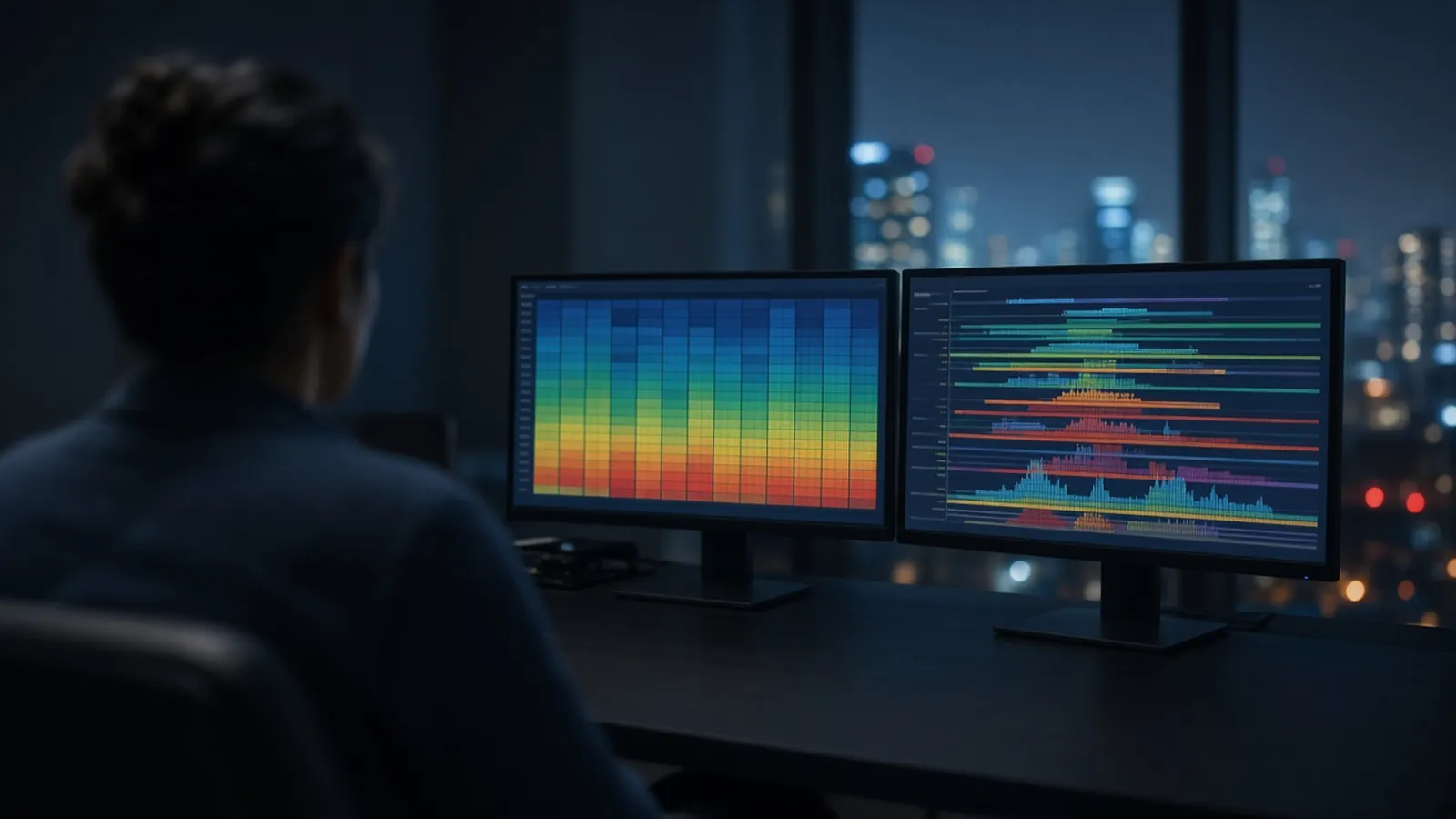
Spatial transcriptomics keeps a tissue address next to an expression measurement. The address might be a spot large enough to hold several cells, a bead, or a coordinate closer to a single cell, depending on the method class. The measurement might be a sequencing library or an imaging-based probe count. The concept is the pairing. Without the address you have bulk expression. Without trustworthy histology you have a library and a photograph that do not describe the same place. This protocol overview is the decision path for that pairing. It is not a vendor recipe, and the times and temperatures stay with the method instructions for the tissue in your cryostat. The non-spatial path is from cells to a gene expression result.
Who needs a coordinate
Use a spatial method when location is the claim: a transcript enriched in a layer, a margin, a structure you can point to on the section. If you only need the average of a dissected region, careful dissection plus bulk RNA-seq is simpler and often better replicated. If you need cell identity and can sacrifice the map, the dissociation route in single-cell RNA-seq in outline is the comparison. The spatial transcriptomics enquiry reference is where a spatial method can be discussed as an enquiry. Read it as a prompt for the specification, not as a booking.
What the coordinate is allowed to mean
Method classes differ in the size of the address they return. Spot-capture and barcoded-array approaches bind RNA from a small area of the section onto a barcoded feature. If that area covers several cells, the count is a mixture. You can still see layers, tumours against stroma, and large gradients. You cannot honestly say the transcript was inside one cell of a named type unless a later analysis, with its own assumptions, tries to unmix the spot. Imaging-based and in situ sequencing approaches hybridise or sequence probes in place and can reach cellular or finer coordinates, usually for a panel or a different read budget. Resolution is chosen with the method. It is not improved by zooming a figure.
Sequencing-based spatial libraries are still RNA-seq libraries. Read structure, depth and strandedness follow from the kit class. A public orientation to short-read sequencing chemistry is the Illumina sequencing overview. It does not define your spot size. Say the spot or cell scale in the same sentence as the gene you plot.
Section quality is the first reagent
The section is the sample. Thickness, freezing, folds, tears and the plane of cutting decide whether a biologist can recognise the tissue. A section that has dried, or that was crushed by the blade, will still produce counts. The counts will not sit on the anatomy you meant. Practise the plane on spare blocks. Stain a serial section, or the same section if the method allows, so the expression overlay has a landmark.
RNA in a section is as fragile as RNA in a tube. The habits in protecting RNA during extraction apply before the slide ever meets a permeabilization buffer: cold handling, a limit on time at ambient temperature, and a record of how the block was frozen. RNase contamination on a brush or a slide is enough to open transcripts in the place you then call a biological dead zone.
Permeabilization and the branch when the chemistry fails
Permeabilization lets the capture chemistry meet the RNA, or lets probes enter the cell, according to the method. Tissues differ. A time that suits a soft mouse brain can under-open a fibrous human specimen, or over-open a delicate one. The manufacturer's protocol for that method class is the source of the time and temperature window. A spare section used as a permeabilization series is the local test.
Read the failure in the image. Weak signal everywhere, with morphology intact, suggests the RNA never reached the chemistry: under-permeabilization, a block that was already degraded, or a reagent missed. Signal that blooms past anatomical borders suggests diffusion: too long, too warm, or a tissue that releases RNA readily. Redo the series. Do not interpret a halo around a structure as a secreted-RNA story until diffusion is ruled out. Saturated signal on a few spots can be a tissue fold, where two layers of cells contributed to one coordinate. Mask folds rather than calling them a niche.
After a sequencing spatial run, the data are counts per coordinate. They still need a differential question: which regions, defined how, compared within which donors. A pattern on one section is a case. Independent animals or donors are the replicates that let you talk about the pattern as repeatable. Public datasets in the European Nucleotide Archive illustrate how variable the metadata for spatial studies can be. Your own record should name the method class, the resolution, the section thickness range you used, and the permeabilization choice.
| Decision | What you are committing to | Branch when it fails |
|---|---|---|
| Region-scale question | A spot or area mixture is acceptable | Do not phrase results as single-cell events |
| Cell-scale question | A method class whose coordinate is cellular | A multi-cell spot will not answer it by relabelling |
| Section readable | Landmarks match the expression overlay | Recut; do not analyse a torn or folded map |
| Permeabilization in range | Signal follows anatomy | Shorten if halos appear; extend if the section is dark |
| One block, one donor | A case illustration | Collect independent donors before a general claim |
| Research coordinate | A hypothesis about place | Keep diagnostic language out of the caption |
A spatial pattern that is really a handling pattern
Edge effects are common. The outside of a section can dry or permeabilize differently from the centre, drawing a rim of signal that looks like a biological margin. Compare with morphology. A crack from freezing can align with a false boundary. A batch of slides processed on a different day can differ in background. If all "induced" regions are the slides from Thursday, you have a batch, and the same caution as any other expression study applies.
Genes that look spatially restricted should be checked for a simple composition story. A transcript expressed in every cell of one type will map wherever that type sits. That is still a spatial fact, and it is not evidence that the neighbourhood induced the gene. Unmixing and cell-type maps are models with parameters. Show the parameter and the marker evidence.
Research coordinates are not a diagnosis
A spatial dataset can be beautiful and still be a research observation on the blocks you cut. It does not diagnose disease, stage a tumour, or prescribe a treatment. Human tissue needs the ethics approval and the biosafety decision of the institution that collected it. The WHO laboratory biosafety manual is general laboratory biosafety guidance, not a permit. Fixatives, stains and permeabilization buffers have hazard notes. Follow the note for the chemical you open. Cryostat blades are a physical hazard of their own. This overview does not set a containment level.
Humidity, frost, and the slide
In a warm, humid laboratory, a section warms and collects moisture the moment it leaves a cold block. Frost on a poorly stored slide becomes water, and water is where RNA diffuses before you meant it to. Keep the cold chain from block to capture as the method instructs, limit how long a slide sits in room air, and write down a power cut that warmed the cryostat or the slide box. A humid afternoon is a sufficient reason for a rim of background. The fix is a repeat section under control, not a stronger colour scale on the damaged map.
What the enquiry has to specify
Name the tissue, the species, whether blocks are already frozen, the resolution the question needs, how many independent donors you have, and what a successful histology image looks like to you. Catalogue classes for sequencing sit in the genomics and sequencing catalogue. The broader sample path is the nucleic acid analysis pathway.
The spatial transcriptomics enquiry reference is the enquiry reference when the coordinate is the point. The mRNA sequencing enquiry reference and the differential expression analysis enquiry reference cover the bulk alternatives and the contrast you may still want. Put the tissue, the resolution and the donor count on the quote request, and ask whether a quotation is possible. A spatial method can be discussed from that specification. The section quality and the claim's resolution stay visible in whatever you write next.
Decide whether a spatial measurement matches the question
- 01Write the coordinate you actually needSay whether the biology lives in a tissue region, in a cell, or in a subcellular compartment. A spot that covers many cells can answer a region question and will blur a cell-to-cell question. Pick the method class whose resolution matches that sentence.
- 02Judge the section before you judge the genesRequire a section with the histology you can interpret: identifiable structures, limited folds, and RNA that has not diffused into a smear. If the morphology is unreadable, stop and cut again. Expression without a trustworthy coordinate is a bulk measurement with extra steps.
- 03Treat permeabilization as a branch, not a defaultFollow the method's own time and temperature guidance for that tissue. Too little permeabilization leaves RNA in the section and yields a weak library. Too much lets RNA wander, so the coordinate no longer matches the cell that made the transcript. A tissue optimisation on a spare block is the usual way to see which failure you have.
- 04Keep the claim inside the resolution and the donorsReport the spot or cell resolution, the section quality, and how many independent donors or animals the pattern was seen in. A beautiful section from one block is one block. It is a research observation, and it is not a diagnosis.
Questions from the bench
Does every spatial method see single cells?
No. Resolution is a property of the method class. Some approaches capture RNA on spots or beads that each mix several cells. Others, including imaging-based in situ methods, can approach cellular or subcellular coordinates. Read the coordinate size before you interpret a marker as belonging to one cell. A spot count is an aggregate at the scale of that spot.
What does permeabilization change?
It opens the section so transcripts can reach the capture chemistry, or so probes can reach the transcripts, depending on the method. The useful window is tissue-specific and is set by the protocol you are following. Under-permeabilized sections look empty. Over-permeabilized sections look blurry, because RNA has moved away from its cellular address.
Can a spatial map replace a pathologist's diagnosis?
Not on the strength of this page. A research spatial dataset can be consistent with a histological pattern and still sit outside diagnostic practice. Diagnosis needs a validated test and the clinical framework that applies where you work. Keep research coordinates and medical reports in separate sentences.
How is this different from dissociating the tissue for single-cell RNA-seq?
Dissociation destroys the arrangement of cells in order to barcode them one by one. Spatial methods try to keep a coordinate, at a resolution the chemistry allows. You lose the map in one case and you may mix cells inside a spot in the other. The outline of the dissociation route is in the single-cell explainer. Choose the loss your question can tolerate.
References
Manufacturer names identify published method classes. Trademarks remain with their owners. Catalogue records on this site are independent references for enquiry. They are not a statement of inventory, distribution rights or a supply commitment. This page is educational. It is not medical advice, a diagnostic protocol or a biosafety approval.
Catalogue
Related products and categories
These links follow the subject of the article into published manufacturer references. A listing is a reference for an enquiry, not a statement of stock or distribution rights.
Continue in this cluster
Related reading
From cells to a gene expression resultHow a laboratory goes from cells to a gene expression result, and how RNA quality decides between a focused RT-qPCR assay and RNA-seq.Single-cell RNA-seq in outlineWhat single-cell RNA-seq adds beyond a bulk average: droplets or wells, empty droplets, doublets, UMIs, sparse counts, and clustering as a parameter.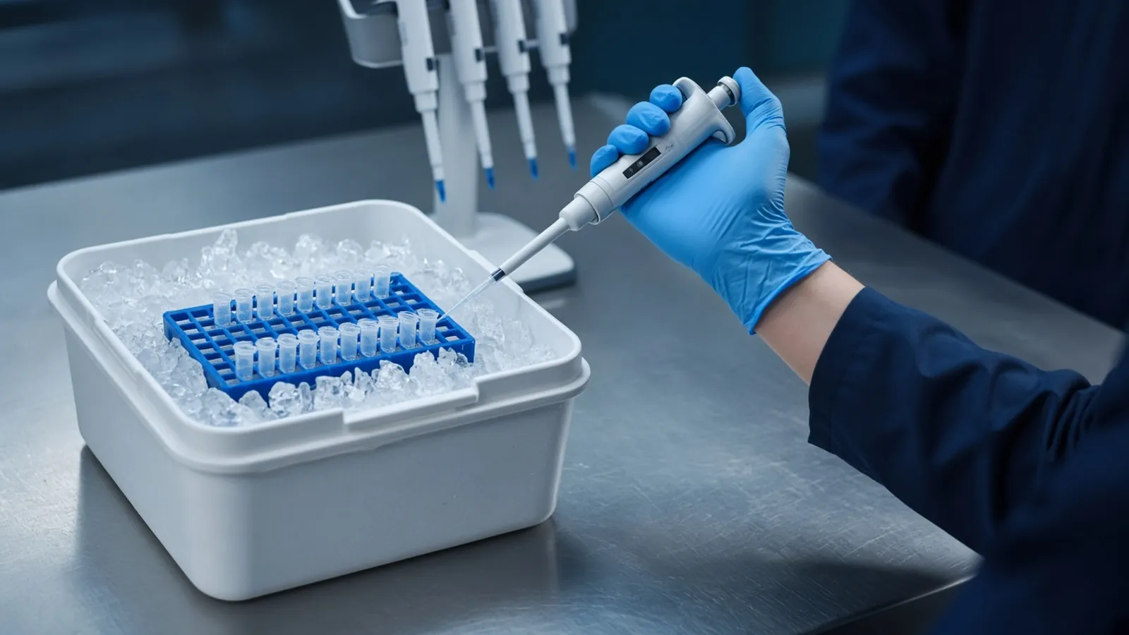 Protecting RNA during extractionHow to keep RNA intact from lysis to elution: RNase control, what A260 ratios do not prove, and the checks that show whether the preparation is still usable.