
guide
Aggregation and cloudy protein samples
Cloudiness, a spin pellet or a void-volume peak means aggregated protein. Do not treat a turbid sample as a concentration or load it onto a column.
- Author
- EVRINTH Editorial Team
- Published
- 8 October 2026
- Updated
- 8 October 2026
- Reading time
- 7 min
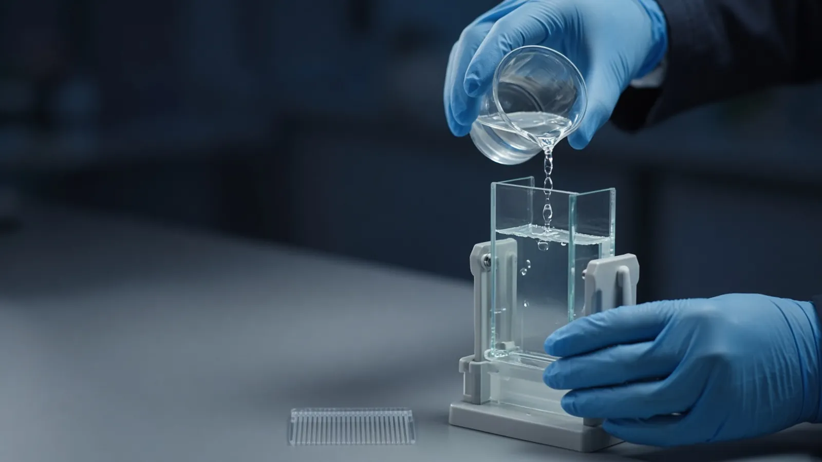
A protein solution that scatters light has stopped being a simple solution, even if a spectrophotometer still prints a number. Cloudiness, a pellet after a modest spin, a smear that never enters a gel, or a peak in the void of a size-exclusion column are all ways of seeing the same class of event: polypeptide that has associated into something much larger than the folded monomer. This guide is for the person about to quote a concentration, load a column, or decide that an expression failed. Expression pellets are compared in inclusion bodies and refolding as a concept. Turning signal into a number is covered in how laboratories estimate protein concentration. What the lane can honestly show is in reading a protein gel.
What the eye, the spin and the column agree on
Hold the tube to the light. Opalescence and white flakes are aggregates or precipitated protein until proven otherwise. A short spin, at a speed your protocol uses to clarify rather than to pellet ribosomes from a dilute buffer, drops much of that solid. If the pellet is large and the supernatant clears, you have separated the problem from the soluble fraction. If the supernatant stays cloudy, the particles are too small to have pelleted or the spin was too gentle. Try the clarification the protocol allows, and do not call the haze "fully soluble".
On a gel, huge assemblies sit in the well. SDS and reductant sometimes dissolve them into the monomer, which tells you the association was not a permanent covalent cross-link. If they remain in the well even after a proper denaturing, reducing load, think about cross-links or about a pellet that was never actually solubilised in sample buffer. Run the supernatant and the pellet in neighbouring lanes. The comparison is more informative than a single lane of the mixture.
Size exclusion tells the same story in solution. Aggregates elute at the void volume, ahead of the folded protein. A void peak plus a monomer peak is a mixture. Injecting that mixture again does not make the void peak go away. It can spread aggregates further onto the bed. Clarify, and if the void peak returns immediately, the column buffer itself is causing the association.
Causes you can actually test
Concentration is the first. Many proteins are soluble at modest levels and cloudy when a spin concentrator pushes them too far. Back-dilute and see whether the haze clears. If it does, the storage concentration was the variable. pH near the isoelectric point removes net charge and often lowers solubility. You do not need a precise pI calculation to test a step of half a pH unit on either side, inside the range the protein's activity tolerates. Salt works both ways. Too little salt lets some proteins stick together. Too much salt salts some of them out. Change one of those variables at a time.
Freeze-thaw is a mechanical cause. Ice formation concentrates solutes in the remaining liquid and then thaws them back. Repeated cycles make this worse. A tube that was clear going into the freezer and cloudy coming out has told you something about storage, not about the gene. Hydrophobic patches, exposed in a partial unfold or by a missing partner, produce a related haze that may not reverse on dilution. A detergent or another additive sometimes belongs in that experiment. It belongs only if the next assay can tolerate it.
None of these tests is a universal cure. They are branch points. If dilution, a pH step and added salt all leave the pellet intact, the soluble yield is whatever remains in the supernatant. Report that. Planning a higher expression of an already insoluble product will make more pellet.
Columns and assays that should not see the solid
Filter or spin before a chromatography injection. A plugged column is an expensive way to learn that flakes do not enter a frit. If you must filter, choose a pore the protocol recommends and accept that protein stuck to the membrane is lost. Rinse only as the protocol allows. Do not scrape a filter cake back into the sample and call it recovered.
Do not report a concentration for a cloudy sample as if it were a solution. Bradford dye and BCA reagents also see scattered light and uneven suspensions. Mix, clarify, then assay the liquid. An A280 that climbs toward shorter wavelengths is scatter until a blank and a clarification prove otherwise. The number you want is the soluble protein you will pipette into the next step.
| Observation | Likely meaning | Next check |
|---|---|---|
| Clears on dilution | Concentration-dependent association | Store more dilute, remeasure |
| Clears away from one pH | Solubility near that pH | Confirm activity in the new buffer |
| Pellet after freeze-thaw | Storage damage | Fresh aliquot, fewer cycles |
| Stays in the gel well after SDS | Very large or cross-linked material | Supernatant versus pellet lanes |
| Void peak on a column | Aggregates in the injected sample | Clarify, then inspect the monomer peak |
| High A280 while cloudy | Scatter added to absorbance | Read only the cleared liquid |
Failure modes that waste a week
Averaging a turbid reading into a yield inflates how much soluble protein you have. Downstream assays then look "less active" per milligram because the milligrams were partly solid. Clarify and recalculate specific activity on the soluble mass. A column back-pressure spike is the same solid announcing itself. Stop. Do not raise the flow to push through.
Reversing aggregation is sometimes possible and often partial. Dilution, a pH move, or fresh reductant for a cysteine-driven tangle are fair tests. Sonication of a precipitated pure protein is a good way to warm and foam it, and a poor way to refold it. If the scientific goal is a soluble active protein, an inclusion-body refolding plan or a new expression condition may beat stirring the same pellet. If the goal is a denatured gel sample, sample buffer and heat are enough. Do not pretend those are the same goal.
Safety and warm rooms
Spins, filters and buffers are routine. The protein source may not be. Biosafety stays with your institution. This is research handling advice, not a clinical product specification.
Warm benches speed unfolding and microbial growth in a salty dilute protein. A sample that was clarified in the morning can opalesce by evening in a hot laboratory. Keep it at the temperature the protein tolerates, and do not leave a concentrator on a sunlit bench. Power cuts thaw freezers and produce exactly the freeze-thaw haze this page is about. Treat every thawed aliquot as a new observation. Spin it before you trust the label concentration. Humidity does not dissolve aggregates, but wet air does ruin hygroscopic buffer salts used to rescue solubility. Weigh those salts properly and check pH.
What to say in an enquiry
Describe the protein mass, the buffer, the concentration where cloudiness appeared, and whether a spin cleared it. Say if a column void peak or a well-bound gel band was seen. Reagents and chemicals is the catalogue category for buffer components you may need to change. Send the observations with a quote. A wider expression question can be discussed from the custom protein expression and purification enquiry reference. That discussion is not a statement that expression, purification or refolding is operated as a service.
Decide what a cloudy protein sample is telling you
- 01Record the look of the sample and the bufferNote cloudiness, flakes or a skin on the liquid, and write the pH, salt and any recent freeze. The description is the observation. A concentration number is not a substitute for it.
- 02Separate solid from liquid before you measureSpin the sample, or filter it if a protocol says a filter is appropriate, and keep both the supernatant and the pellet. A gel of each fraction shows whether the protein you care about stayed soluble.
- 03Do not load the suspension onto a columnAggregates plug resin and appear in the void of a size-exclusion run. Clarify first. If the clarified sample still clouds as it enters the column buffer, stop and change conditions rather than forcing the injection.
- 04Quantify only the fraction you will useRead the clear supernatant with an assay the buffer allows. Scatter makes a turbid A280 look concentrated. Report the soluble measurement, and say that the pellet was removed.
Questions from the bench
Is a cloudy protein the same thing as an inclusion body?
Not necessarily. An inclusion body is insoluble material from expression, packed inside the cell before lysis. Cloudiness in a purified tube can come from later aggregation: concentration, pH, freeze-thaw, low salt or a hydrophobic surface. The handling is related. The history is different.
Why does absorbance rise when the sample turns cloudy?
Particles scatter light, and scatter adds to the apparent absorbance, especially toward shorter wavelengths. The spectrophotometer is no longer reporting only tryptophan and tyrosine. Clarify the sample and blank properly before you convert the reading into a concentration.
What does material in the gel well usually mean?
Large aggregates often fail to enter the resolving gel and sit in the well or the stacking gel. A reducing, SDS-denatured sample that still will not enter may be cross-linked or badly precipitated. Compare a spun supernatant with the resuspended pellet before you decide the protein vanished.
Should I filter every protein before a column?
Clarify anything that is visibly cloudy or that has been frozen and thawed. A short spin is often enough for a small prep. A filter is appropriate when the protocol and the protein tolerate it. Forcing a thick precipitate through a fine filter mostly blocks the filter. It does not redissolve the solid.
References
Manufacturer names identify published method classes. Trademarks remain with their owners. Catalogue records on this site are independent references for enquiry. They are not a statement of inventory, distribution rights or a supply commitment. This page is educational. It is not medical advice, a diagnostic protocol or a biosafety approval.
Continue in this cluster
Related reading
Reading a protein gelHow to read a protein gel: what SDS-PAGE bands, smears, ladders and loading differences can support, and what they cannot identify.Inclusion bodies and refolding as a conceptAn inclusion body is insoluble expression, not proof a protein can never fold. Refolding is empirical, and activity rather than a band shows success. How laboratories estimate protein concentrationHow laboratories estimate protein concentration with absorbance, dye-binding and copper assays, and why the number depends on the standard.