
comparison
Inclusion bodies and refolding as a concept
An inclusion body is insoluble expression, not proof a protein can never fold. Refolding is empirical, and activity rather than a band shows success.
- Author
- EVRINTH Editorial Team
- Published
- 8 October 2026
- Updated
- 8 October 2026
- Reading time
- 7 min
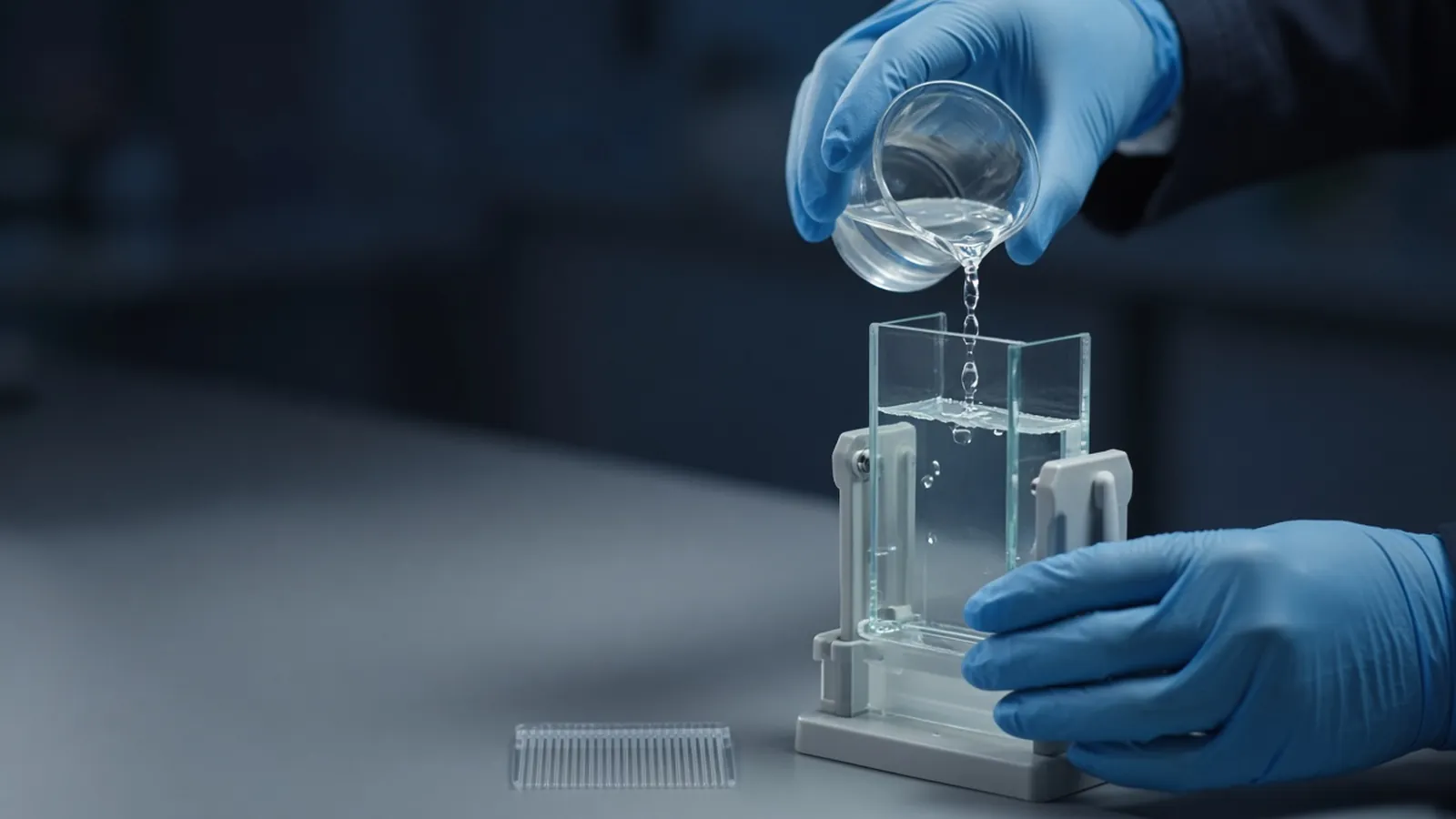
Escherichia coli sometimes answers an expression plasmid by packing the polypeptide into a dense insoluble lump. That lump is an inclusion body. It is evidence of aggregated expression, not a proof that the chain is incapable of a native fold. The comparison this page supports is between chasing solubility during expression and accepting the insoluble material, then trying to unfold and refold it. Both routes need a gel, described in reading a protein gel, and a functional test. Capture after either route is a chromatography problem, outlined in affinity and ion-exchange protein purification. Cloudiness later, in a bottle you thought was folded, is a different event and is covered with aggregation and cloudy protein samples.
What the pellet is
After lysis, a spin separates soluble protein from debris and from inclusion bodies. Washing the pellet, often with a detergent-containing buffer, removes membrane fragments and some stuck contaminants. The wash is not the refolding. A Coomassie gel of the washed pellet that shows one dominant band at the expected mass is encouraging enrichment. It still does not identify the protein, and it says nothing about activity. Host proteins can remain. Quantification of a suspension, or of a pellet resuspended by wishful mixing, is not a concentration. Dissolve first, then measure by a method the solvent allows.
Inclusion bodies form for dull reasons as well as interesting ones: very high expression, a temperature that is too high for that chain, missing partners, disulfides that cannot form in the reducing cytosol, or a construct that is simply unhappy. Turning the inducer down or dropping the temperature is part of the comparison, not a footnote. Addgene's protocol collection is a public place to see how expression notes are usually structured. Copy the decision logic, not a fixed induction table.
Solubilise, then take the denaturant away
Urea and guanidine hydrochloride are the denaturant classes most often used to dissolve washed inclusion bodies. They unfold the chain and bring it into solution. Typical practice uses high molarities, the sort of multi-molar concentrations textbooks associate with these reagents, for long enough that the pellet clarifies. The exact concentration and time belong to the protocol for that protein. Urea and guanidine are not drop-in substitutes. Guanidine is ionic. Urea solutions can accumulate cyanate, especially when warm and aged, and cyanate can carbamylate proteins. Prepare urea with the care the protocol states, and do not store a warm stock for convenience.
Refolding means removing the denaturant so the chain can seek a soluble structure. Dilution into a large volume, dialysis, or an on-column removal are the usual classes. All of them can precipitate. A screen of pH, salt, redox additives for disulfide proteins, and additives such as arginine is empirical. There is no sequence feature that lets you skip the screen and declare a buffer in advance. Rapid removal often crashes the protein. Very slow removal can also produce aggregates. The outcome is judged on the soluble fraction after a spin, and then on function.
Activity is the result that matters
A clear supernatant and a single band mean you have soluble polypeptide. They do not mean you have the enzyme, the binder, or the correctly paired disulfides. Compare specific activity, or a binding signal per mass, with a preparation that was soluble from the start if you have one. If you do not, define the functional assay before you celebrate the gel. A refold that is soluble and inactive may be misfolded, inhibited by leftover denaturant, or missing a cofactor. Those are different next steps. Residual urea or guanidine can also suppress an assay, so buffer-exchange a sample before you call the activity zero.
Many proteins should never enter this path. If a homologue is known to express in a soluble fraction under milder conditions, try that comparison early. Disulfide-rich proteins may need a host or compartment that can oxidise them, rather than a heroic refold from the cytosol. A fusion partner that improves solubility is another legitimate arm of the comparison. It adds mass and may need removal. Put that cleavage on the plan before you choose the fusion for convenience.
| Route | What you can gain | What still has to be shown |
|---|---|---|
| Soluble expression | Folded material without a denaturant step | That the band is the intended product and is active |
| Washed inclusion body | Enrichment of insoluble product | Identity, then a solubilisation that is real |
| Denaturant plus refold | A second chance at a soluble chain | Activity or binding after denaturant removal |
| Different host or fusion | A way around the pellet | The fusion or host did not create a new artefact |
Failure modes worth separating
Precipitation during the removal step means the conditions, the concentration, or the speed of removal were wrong for that chain. Lower the protein concentration and change one variable. A soluble protein with no activity and a normal gel needs the assay checked for leftover denaturant, missing cofactor and pH before you condemn the fold. A gel full of fragments means proteolysis during the long refold. Add the inhibitor classes your protein can tolerate, and shorten the time. A beautiful band that is the wrong mass is still the wrong protein. Check the construct against UniProt or your translated plasmid, including tags.
Do not call a cloudy refold a concentration. Clarify it, measure the supernatant, and look at the pellet on a gel. If most of the stain is in the pellet, the soluble yield is the small number, not the absorbance of the suspension.
A redox pair is a separate branch for proteins that need disulfides. Adding a thiol and a disulfide reagent, in a ratio taken from a protocol rather than from guesswork, can allow pairing during the removal of denaturant. The gel under non-reducing conditions then becomes useful: a ladder of mis-paired species is a failed screen even if the reducing lane looks like a single subunit. Keep that redox experiment distinct from a plain dilution screen so you know which change helped.
Safety and the room
Urea, guanidine and the reducing or oxidising additives used in disulfide screens are chemical hazards. Follow the protocol's handling notes. Concentrated denaturants are not drain-casual in a teaching sense: dispose of them as your institution requires. This page is research education. It is not a manufacturing method and not biosafety approval for the strain you are lysing.
Refolds that sit for many hours in a warm laboratory grow microbes and drift. A cold room that is not actually cold after a power cut is how a slow dialysis becomes a spoiled one. Record the temperature you achieved. Cyanate risk in urea climbs when solutions sit warm. Make the denaturant when you need it, with the quality the protocol asks for.
What to send if you want the route discussed
An enquiry is more useful when it says whether solubility was already tried, what the pellet gel looked like, whether disulfides are expected, and which activity would count as success. Buffer salts and denaturant classes sit with reagents and chemicals. Put the scientific constraints in a quote. The custom protein expression and purification page is an enquiry reference for discussing an expression route. It is not a claim that proteins are expressed or refolded as an operated service.
Questions from the bench
Does a white pellet after lysis mean the protein can never fold?
No. It means that under those expression conditions a large share of the polypeptide was insoluble. Many proteins that form inclusion bodies can fold when they are expressed more slowly, in another host, or after a deliberate refolding screen. The pellet is a result, not a law.
What is the difference between urea and guanidine here?
Both are denaturant classes used to solubilise inclusion bodies. They are not interchangeable stocks. Guanidine is a salt as well as a denaturant, and urea solutions can develop cyanate that modifies proteins if they are old and warm. Follow a protocol for the denaturant you chose, and do not copy molarities from the other one.
Why is a band after refolding not enough?
A gel shows that polypeptide of about the right mass is back in the soluble fraction. It does not show that the active site, the disulfide set, or the oligomer is correct. Activity, a binding measurement, or another functional assay is the success criterion. A soluble inactive protein is a completed failure mode, not a yield.
Should every difficult protein be refolded from E. coli?
No. Refolding is empirical and can consume more effort than changing the expression route. Lower temperature, a different fusion, or a host that supports disulfides or solubility is often the better comparison. Choose refolding when the insoluble route is justified, not as a default.
References
Manufacturer names identify published method classes. Trademarks remain with their owners. Catalogue records on this site are independent references for enquiry. They are not a statement of inventory, distribution rights or a supply commitment. This page is educational. It is not medical advice, a diagnostic protocol or a biosafety approval.
Catalogue
Related products and categories
These links follow the subject of the article into published manufacturer references. A listing is a reference for an enquiry, not a statement of stock or distribution rights.
Continue in this cluster
Related reading
Reading a protein gelHow to read a protein gel: what SDS-PAGE bands, smears, ladders and loading differences can support, and what they cannot identify.Aggregation and cloudy protein samplesCloudiness, a spin pellet or a void-volume peak means aggregated protein. Do not treat a turbid sample as a concentration or load it onto a column. Affinity and ion-exchange protein purificationHow affinity capture and ion exchange pull a protein from a lysate, and why each collected fraction still needs an identity check.