
troubleshooting
Blocking and plate washers
How blocking reagents and plate washers change ELISA background, and how to tell a sticky well from a wash that never finished.
- Author
- EVRINTH Editorial Team
- Published
- 8 October 2026
- Updated
- 8 October 2026
- Reading time
- 9 min
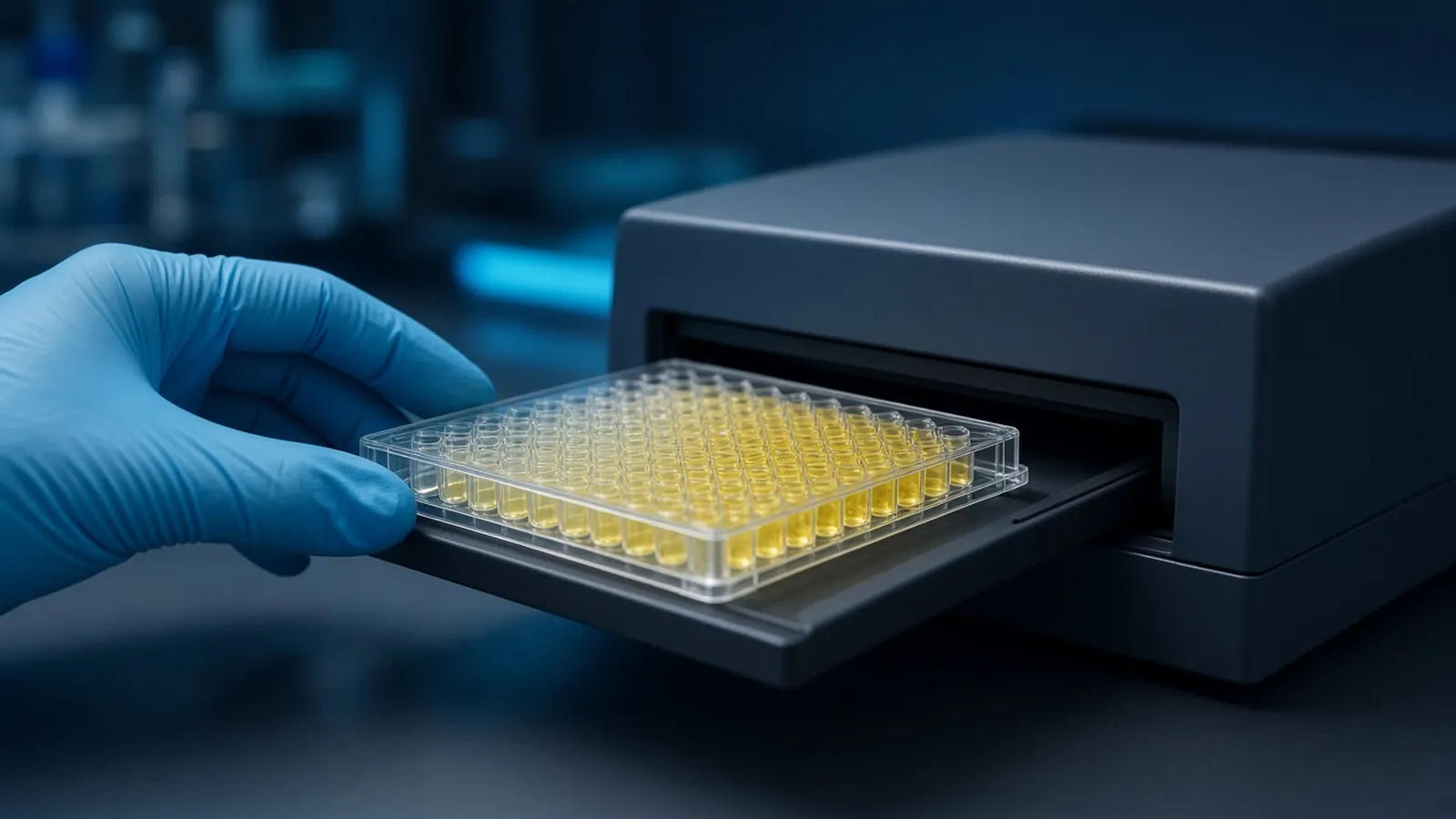
High background and a missing signal are not one troubleshooting tree with two moods. High background means something is sticking that should have been blocked or washed away. Low signal means the sandwich, the enzyme or the substrate never produced colour where a positive control says it should have. Blocking reagent and the plate washer sit on both trees, and they fail in different directions. The assay formats those wells belong to are in ELISA formats, controls and readout. This page is what to change when the plastic or the wash, rather than the biology, is writing the result.
Separate the symptom before you change three reagents
Start from the blank, not from the most interesting sample. A zero-analyte well that is almost as dark as the mid standard is a background problem. A positive control that matches the blank, on a plate whose substrate still turns over in a test tube with fresh enzyme, is a signal problem. If both are true, fix background first. You cannot judge a rescue of the signal while every well is already painted.
Write down whether the pattern is the whole plate, a column, an edge, or a single well. Whole-plate background points at the blocker, the detection antibody concentration, or a wash buffer that every well saw. A column points at a washer pin. An edge points at drying or temperature. A single well points at a splash or a bubble. The pattern is the hypothesis.
What the blocker is occupying
After the capture antibody or the antigen is coated, a large fraction of the plastic is still bare. Detection antibody, enzyme conjugate and even the analyte will adsorb to that bare polystyrene and create signal that has nothing to do with a sandwich. A blocking solution fills those sites with something you hope the detection system will ignore.
Common classes are albumin, often bovine serum albumin, casein, non-fat dried milk, serum of a species the secondary does not see, and commercial blockers that are protein or synthetic polymer. Detergent in the wash, commonly a tween class, reduces weak sticky interactions during the washes but is not a substitute for a block. Concentrations and times belong to the protocol you are following. A typical protein block is in the low percent range for half an hour to a few hours. Use the sheet for the product you opened, not a number remembered from a different kit.
The blocker can be the analyte. Do not block an albumin assay with albumin, a casein assay with milk, or a bovine immunoglobulin assay with ordinary bovine serum albumin that still contains immunoglobulin. The zero well will be high and the standard curve will look compressed because you have added analyte to every well including the blank.
The blocker can also be a ligand the detection system sees. Milk and some serum preparations contain biotin, which occupies avidin or streptavidin and either raises background or kills a biotinylated detection layer. Milk contains phosphoproteins that phospho-specific antibodies may bind. Bovine immunoglobulin in milk or crude albumin can be recognised by anti-bovine secondaries and by some anti-goat or anti-sheep reagents that were not fully cross-adsorbed. If the secondary-only well goes dark only when that blocker is used, the blocker is part of the stain. An orientation to why secondary reagents see immunoglobulin is in the molecular-cell background on the NCBI Bookshelf. Your plate still has to show it locally.
What the washer is doing, pin by pin
A wash removes unbound detection reagent from the well volume. It does not remove a reagent that has specifically bound, unless the buffer is harsh enough to strip a weak antibody. Too few cycles leave enzyme behind and the blank rises. Too many cycles, or a buffer with the wrong detergent and salt, can lower a real signal. Record the cycle count, the soak time and the buffer recipe. Change one of those at a time.
Washer hardware fails in three ways that look like biology. Residual buffer left in the well dilutes the substrate or the next antibody, so signal falls, and it can leave a film of detection antibody, so background rises. You see it as a larger coefficient of variation and, often, as bubbles. Clogged or misaligned pins skip a well or a column. That column is either unwashed and dark or unfilled and pale, in register with the manifold. Aspiration that sucks the well dry and then waits lets the coated surface change. Patchy background and an edge that is worse than the centre are the usual picture. A manual wash with a multichannel pipette avoids manifold geometry and introduces its own failure: uneven tip heights, a splash from a forceful dump, and a plate that was tapped dry and then forgotten.
Choose manual washing or a washer as a class you can repeat. This is not a brand ranking. A washer you understand, with a daily water rinse and a dye check, beats an unexamined machine. A careful hand wash beats a washer with a blocked pin. Public wash habits are sketched in places such as protocols.io and in reagent notes such as Promega protocols. Copy the logic, then keep the cycle count that your own blank accepts.
Water quality sits under both methods. Wash buffer made from a poor water grade grows microbes and leaves residues that show up as background. Where laboratory water fails is its own subject in laboratory water types and where they fail. If the blank climbed the week the polishing cartridge was exhausted, believe the calendar.
| What you see | Blocker-sided explanation | Washer-sided explanation |
|---|---|---|
| Every zero well is dark, samples only a little darker | Blocker is the analyte, contains biotin or Ig, or was too thin | Wash cycle count too low for this detection antibody, on every well |
| One column dark on every plate | Unlikely, unless that column received a different block | Clogged, misaligned or unprimed pin |
| Patchy wells, worse after a pause | Block dried and cracked, or was contaminated | Aspiration left wells dry for too long |
| Signal low everywhere, blank quiet | Over-blocking or a blocker that inhibits the enzyme | Too many cycles, or residual buffer diluting the substrate |
| Background climbed only in serum samples | Blocker does not cover sites the serum proteins reveal | Residual sample matrix left in the well and the next reagent bound it |
How to reason from the plate you already have
Do not change the blocker and the washer programme on the same afternoon. You will not know which one saved you.
If the blank is high and the secondary-only well is high, dilute the detection reagent and confirm the block before you buy a new primary antibody. A primary that was never added cannot be the cause of a secondary-only stain. If the blank is high only when sample matrix is present, the matrix contains a sticky protein or an immunoglobulin the secondary sees. More of the same blocker may help, or a different class may help, or the secondary needs to be pre-adsorbed against that species. Extra substrate will not help.
If the blank is quiet and the positive control is quiet, the block may be too heavy, the wash may be stripping the capture coat, or the enzyme may be dead. A substrate-plus-enzyme spike in an empty well separates a dead substrate from a dead sandwich. Residual wash buffer diluting that spike is a washer problem. A spike that colours, beside a sandwich that does not, sends you back to the antibodies.
If a column fails, stop running study plates on that manifold. Push wash buffer through by hand or run a dye plate. A pin that delivers dye to every well can go back into service. A pin that does not is the result you have been calling biology.
Waste, azide and who decides the drain
Wash waste contains diluted antibody, often azide, sample proteins, and detergent. Whether that waste can go to a sink is an institutional decision, not a habit copied from a neighbouring bench. Human-derived samples in the wash make the decision stricter. Stop reagent is often acid and should not be casually combined with azide-containing waste, because some azide and acid mixtures are a chemical hazard. Ask the safety officer. The WHO Laboratory Biosafety Manual is a public reference for the biosafety side of specimen handling. A quieter blank does not turn the plate into a diagnostic test.
Humidity, a sour blocker and the water in the washer lines
Protein blocker left at room temperature in a humid laboratory grows. A casein or milk solution that smells sharp or looks cloudy is not a thrifty reagent. It is a source of background and of proteases. Make the volume you will use, keep it cold, and do not top up yesterday's bottle. In a hot room, a plate that waits for its next reagent dries from the edges inward even if you meant to leave it wet. Work in batches you can actually wash and refill.
Washer bottles of dilute buffer also grow when they sit warm between uses. A line full of biofilm dispenses background. Rinse with the water grade your protocol names at the end of the day, and do not let a manifold dry full of salt. If a power cut stops a washer mid-cycle, those wells are not half-washed in a way you can ignore. Finish the wash by hand or repeat it, and mark the plate. A partial machine cycle is not a standard cycle.
What to specify when you enquire
Name the analyte, so nobody proposes a blocker that is the analyte. Name the detection system, especially biotin-streptavidin or a phospho-specific antibody, and the sample species. Say whether you wash by hand or by manifold, and how many cycles the current blank can tolerate. State the water grade you can actually make. The reagents and chemicals catalogue is a route into buffer and blocker classes. The molecular biology pathway sets the method among others. A blocking scheme can be discussed through the quote request. Ask which blocker classes are incompatible with the detection chemistry. Do not ask a catalogue page to rank washers, and do not expect a reagent line to arrive already shown against your matrix.
Questions from the bench
Why does the blocker sometimes become the thing the assay measures?
Blocking proteins occupy plastic, but they are still proteins. If you block with the analyte, or with a preparation that contains it, every well looks positive. Milk and some serum blockers also carry immunoglobulin or biotin that particular detection systems recognise. The blocker has to be invisible to the antibodies you are using.
What does a clogged washer pin look like on the plate?
It rarely looks random. A column or a single well stays dark, or stays pale, in the same position on every plate that washer touches. Residual buffer in that well dilutes the next reagent or leaves detection antibody behind. Clean or replace the pin, and confirm with a dye-filled test plate before you blame the antibody.
Should wells be left dry after the last wash?
A short interval while you add the next reagent is normal. A plate that sits aspirated and dry for a long time often shows higher, patchier background because the coat and the block have changed. Follow the protocol's instruction on whether to tap the plate dry or to leave wells wet, and do not park a dried plate while you take a break.
Is a manual wash a lesser method than a machine wash?
They are different classes of technique. A careful manual wash can be excellent and slow, and a neglected washer can paint stripes across a study. Choose the class you can perform the same way on every plate you will compare. The choice is not a ranking of brands.
References
Manufacturer names identify published method classes. Trademarks remain with their owners. Catalogue records on this site are independent references for enquiry. They are not a statement of inventory, distribution rights or a supply commitment. This page is educational. It is not medical advice, a diagnostic protocol or a biosafety approval.
Catalogue
Related products and categories
These links follow the subject of the article into published manufacturer references. A listing is a reference for an enquiry, not a statement of stock or distribution rights.
Continue in this cluster
Related reading
ELISA formats controls and readoutELISA formats, controls and readout: how direct, sandwich and competitive assays turn a binding event into a number that stays inside the curve. Laboratory water types and where they failResistivity, organic carbon and endotoxin answer different questions. A water grade fails when the assay cares about a contaminant the meter cannot see.Troubleshooting a high backgroundHigh background means the blank is loud. Separate a dirty blank from a plate that is loud in every well before you subtract and move on.