
glossary
Troubleshooting a high background
High background means the blank is loud. Separate a dirty blank from a plate that is loud in every well before you subtract and move on.
- Author
- EVRINTH Editorial Team
- Published
- 8 October 2026
- Updated
- 8 October 2026
- Reading time
- 9 min
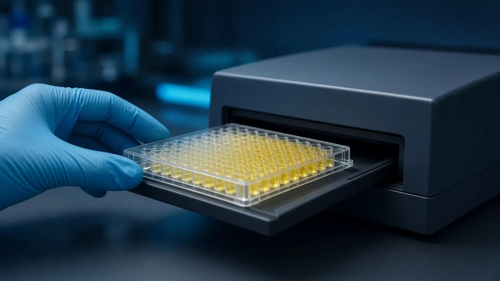
Troubleshooting a high background starts by naming three words that people collapse into one complaint. Background is signal you did not intend to attribute to the analyte. The blank is the well you chose, in writing, to estimate that unintended signal. Nonspecific binding is one physical cause: a detection reagent stuck to plastic, to the blocker, or to something in the sample that is not the analyte. A plate can also go yellow because the enzyme ran too long, which is not binding at all. The formats and the control wells that make this distinction possible are in ELISA formats, controls and readout. This page is a decision tree for a research plate. It is not a clinical interpretation.
Read the pattern before you touch the dilution
Lay the optical densities out in plate order, not as a bar chart of groups. Three patterns cover most loud plates.
The substrate blank is high and so is everything else. The colour reagent was already converting, or it sat on the bench until it did. Discard that substrate. Do not calculate concentrations.
The substrate blank is quiet, and the zero standard, the no-analyte wells, or the no-capture wells are high. Enzyme meets substrate only where conjugate was bound. Something captured the conjugate without your analyte. That is the nonspecific binding branch.
Every sample well is high, the zero wells are high, and a true difference between low and high standards has vanished. Either the curve is saturated because substrate time or conjugate level was excessive, or analyte has contaminated a common reagent so there is no longer a zero. Those two causes ask for different fixes. Look at whether a freshly opened diluent restores a zero before you halve the antibody.
A standard curve that still rises, with a zero that is merely a bit high, is a different problem from a flat loud plate. You may still have a window. You do not widen that window by subtracting a huge blank and calling the remainder a concentration. Store raw values. Fit only if the zero and the standards still form a shape you declared in the protocol.
Washes that never removed the unbound enzyme
Unbound conjugate left in the well will turn substrate. Insufficient wash is the first mechanical cause. Count the washes you actually did, not the number written on an old card. A washer that leaves a visible residual in the corners, or a manual wash that skips the rim of the well, leaves enzyme behind. Extra washes are not free either: a harsh buffer or an excessive cycle can strip a weak capture. Change one thing. If you add washes, keep the antibody dilution constant so you know which change quieted the blank.
Detergent in the wash buffer, often a low level of a non-ionic detergent, is part of many protocols. Too little, and sticky conjugate remains. Too much, or a detergent the capture antibody dislikes, and signal falls everywhere, which is the opposite complaint. Follow the method you are copying, and record the buffer recipe. Preparing that buffer is ordinary bench chemistry: the pH and the water quality decide whether the wash is what you think it is. Preparing a buffer and checking pH is the relevant habit. A wash bottle that has grown a film is a contamination source, not a time-saver.
Blocker omitted, exhausted, or seen by the detection system
If the blocking step was skipped, the plastic is a binding surface for every protein you add afterwards, including the detection antibody. The blank rises because the well itself is the capture. Restore the block and rerun the controls before you redesign the assay.
A blocker that was reused until it smelled or clouded is not a blocker. Proteins spoil. A dried plate, where blocking solution evaporated and left a tide mark, has bare plastic again at the edge of the well. That well will bind conjugate. Keep plates covered and wet for the times the protocol states. If a plate must be stored after coating, use the conditions the coating method allows, not an open stack in a fume hood.
The subtler failure is a blocker the detection system recognises. Biotin in a milk or serum blocker feeds a biotin-avidin or streptavidin amplification and paints every well. Phosphatases or phosphoproteins in milk can create trouble for alkaline phosphatase detection or for phospho-specific antibodies. If the blank fell when you changed blocker class and changed nothing else, you have the cause. Do not describe that plate as high expression.
Primary antibody, secondary antibody, and a conjugate that is too strong
The primary antibody can raise background when it binds the matrix, when it is used far above the dilution that saturates specific binding, or when it contains aggregates that stick. A no-primary well that is quiet, beside sample wells that are loud, points at the primary or at real analyte. A no-primary well that is as loud as the samples points at the secondary or the conjugate.
Secondary antibody that is too concentrated is a classic loud blank. The extra immunoglobulin sticks. Dilute it in a series on a small plate, with the zero standard present, and pick a dilution where the zero is quiet and a positive control still responds. One guessed dilution repeated for months is how a loud plate becomes normal. The conjugate species must still match the primary host. A wrong species usually gives no signal rather than high background, but a dirty conjugate can do both in different hands.
Enzyme conjugates that have been contaminated with analyte, or diluents that were pipetted with a tip that previously touched a standard, abolish the zero. This is easy to do with a multichannel when the standard row is next to the diluent reservoir. Use a dedicated reservoir for the zero and the blanks. If a new aliquot of diluent restores the zero, retire the contaminated one. Do not try to calculate around it.
Substrate left on the bench
Horseradish peroxidase and alkaline phosphatase keep working until substrate is depleted, the product inhibits them, or you stop them as the sheet instructs. A timer that starts late because the first rows were filled slowly means those rows are darker for a reason that has nothing to do with analyte. Start the timer when the chemistry says to start it, and stop the whole plate together if the protocol is an endpoint. Promega protocols illustrate how enzyme sheets state time and temperature. Use the sheet for the substrate you opened.
A warm room shortens the useful window. If the blank is still climbing when you stop, you waited too long for that temperature. Shorten development or lower the conjugate, and say which one you did. Do not subtract the climbing blank and keep a kinetic mess as if it were an endpoint concentration.
A table for where the colour sits
| What is loud | What is quiet | Likely branch | What you change first |
|---|---|---|---|
| Substrate blank and all wells | Nothing | Substrate already converting, or gross contamination of the colour reagent | New substrate. Do not fit a curve |
| Zero standard and no-analyte wells | Substrate blank | Conjugate or detection antibody binding without analyte | Washes, blocker class, secondary dilution |
| No-primary wells | Wells that never received secondary | Secondary or enzyme system | Secondary dilution or a fresh conjugate |
| Every well equally, standards included | No window between low and high standard | Saturation, shared contamination with analyte, or dried plastic | Fresh diluent, shorter development, then a uniformity check |
| Only sample wells, zero stays low | Blank | This may be analyte or cross-reactivity, not background | Do not call it background. Inspect the curve and the antibody |
Stop before a subtraction becomes the result
If the blank optical density is a large fraction of the sample optical density, you do not have a measurement with a small offset. You have two large numbers and a shaky difference. Pipetting error on both wells dominates the difference. Report the plate as failed background, show the raw table, and change the cause. A later, quieter plate is the replicate. The arithmetic on the loud plate is not.
There is a glossary trap in the word corrected. Corrected optical density sounds official. Unless the protocol defines the blank and shows the uncorrected numbers beside it, another person cannot audit the correction. Keep the phrase out of the claim sentence until both columns exist.
Plate maps and worked examples on protocols.io are useful when you design the control wells. The Addgene molecular biology reference is a general bench reference, not an ELISA kit insert. Neither source can see your washer alignment.
Safety while you are rerunning the plate
Stop solutions are often strong acids. Substrate components can be irritants. Antibody stocks may contain sodium azide or other preservatives. Do not tip a loud plate down a sink because you are annoyed with it. Follow the chemical and biological waste rules of the institution. Specimens that may contain pathogens stay inside the biosafety decision already made for that work. The WHO Laboratory Biosafety Manual is a public reference for building that decision. A high blank is still a research observation. It does not diagnose anyone.
A warm afternoon that develops every well
Colour development is faster when the room is hot. An incubation that was acceptable in a cooled morning can push the blank up in an uncooled afternoon, even though the pipette work was identical. Note the room temperature next to the development time. If you cannot hold the substrate step at the temperature the sheet asks for, shorten the time until the blank behaves, and do not compare that plate with a colder one as if the clocks were equal.
Wash buffer left open overnight in a warm, humid laboratory can grow microbes that later stick or that spoil the detergent. Make the volume you need, label the date, and discard on the schedule you set. A power cut that stops a plate washer mid-cycle leaves some columns unwashed. Those columns will be darker. Do not average them into the washed columns. Finish or repeat the wash when power returns, and record the split.
What to say when you ask for help with a loud plate
Describe the pattern from the table: which wells are loud, which are quiet, the blocker class, the conjugate and its dilution, the substrate time and wavelength, and whether the plate dried. Those facts let a reagent class be discussed. The reagents and chemicals catalogue is a place to look at buffer and reagent classes. The molecular biology pathway is the surrounding methods context. Use the quote request to ask whether a quotation is possible. A method can be discussed from the pattern you send. Ask for the blocker compatibility and the suggested conjugate range in the reply, and keep the raw optical densities so a suggestion can be tested rather than believed.
Questions from the bench
Can I subtract a high blank and keep the sample values?
Only when the blank is small beside the signal and you have already stored the raw optical densities. A blank that sits near the samples means the assay has no window. Subtraction then invents differences out of noise and pipetting error. Fix the blank before you interpret a remainder.
Why would only the zero standard be high while the substrate blank is quiet?
The enzyme and the colour reagent are behaving. Something in the full protocol is sticking: the detection antibody, the conjugate, or analyte that has contaminated a buffer. A quiet substrate blank rules out a substrate that had already turned. It does not rule out nonspecific binding.
Does a darker blocker always mean a quieter plate?
More protein is not automatically a better block. The wrong protein can be seen by the detection system, as when a biotin-rich blocker meets an avidin conjugate, or a phospho-protein-rich milk meets a phospho-specific reagent. Match the blocker class to the chemistry, then judge it by the blank.
The whole plate, including a spiked control, is equally yellow. What failed?
Equal colour in every well usually means a step that does not depend on analyte: substrate left too long, a conjugate dumped into the substrate by a poorly rinsed tip, or a plate that dried and then bound detection reagent everywhere. The standard curve cannot be fitted honestly to a flat high line.
References
Manufacturer names identify published method classes. Trademarks remain with their owners. Catalogue records on this site are independent references for enquiry. They are not a statement of inventory, distribution rights or a supply commitment. This page is educational. It is not medical advice, a diagnostic protocol or a biosafety approval.
Catalogue
Related products and categories
These links follow the subject of the article into published manufacturer references. A listing is a reference for an enquiry, not a statement of stock or distribution rights.
Continue in this cluster
Related reading
ELISA formats controls and readoutELISA formats, controls and readout: how direct, sandwich and competitive assays turn a binding event into a number that stays inside the curve. Preparing a buffer and checking pHPrepare a buffer at a stated pH and temperature, record the salt form, and treat a drifting electrode or a bad slope as a failed control.Troubleshooting no signalA flat research ELISA is a broken chain, from coating to wavelength. Separate a truly blank plate from a signal that merely sits below the curve.