
application
Troubleshooting no signal
A flat research ELISA is a broken chain, from coating to wavelength. Separate a truly blank plate from a signal that merely sits below the curve.
- Author
- EVRINTH Editorial Team
- Published
- 8 October 2026
- Updated
- 8 October 2026
- Reading time
- 11 min
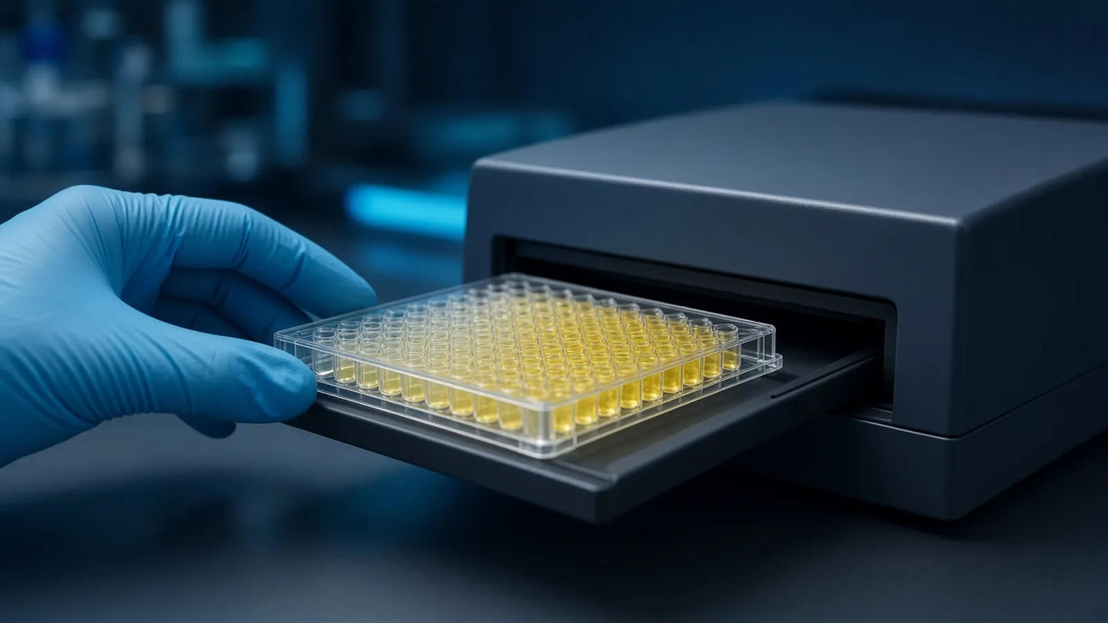
Troubleshooting no signal, in this application, means a research ELISA that came off the reader as a flat line. The wells look like the instrument baseline. Before anyone writes that the treatment abolished the analyte, the chain from coating to wavelength has to be walked. A flat plate is often a reagent that never met its partner. The control logic for a working plate is in ELISA formats, controls and readout. Checking that the primary antibody can see the target at all is in choosing and checking a primary antibody. This is not a guide to clinical false negatives. A diagnostic miss is a different claim, under a different quality system.
What the flat file is allowed to mean
Open the raw optical densities, not a spreadsheet that has already subtracted a blank and hidden the zeros. If every well, including the highest standard and a positive control, sits on the instrument baseline, you do not have low biology. You have an assay that did not transduce binding into light or colour. If the standards rise and the samples sit with the zero, you may have low samples, a matrix that hides analyte, or a hook effect at the other extreme. Those are interpreted only after the standards have proved the chain works. Rank nothing on a plate whose calibrators failed.
Note the reader error flags. A wavelength that was left on the previous user's setting, a lamp that did not strike, or a plate that was unread because the drawer did not close, all produce empty-looking files. Confirm a physical read of a coloured well, even a spare well you tint with a drop of stopped substrate, before you throw away the plate. The photograph of a developed plate entering the drawer is the measurement step. It can only report what the optics are set to collect.
Was capture on the plastic at all
Coating fails quietly. The capture antibody may have been diluted into the wrong buffer, left at a temperature the sheet forbids, or pipetted into a plate type that does not bind protein. Some plates are treated for cell culture and are poor at adsorbing immunoglobulin. Some are sold pre-blocked. Read the plate product page before you blame the antibody. A coating incubation that dried out leaves a rim of antibody and a bare centre, which can look like weak signal rather than a true zero. If the entire plate is zero, drying is less likely than a missed reagent.
A useful branch is a well spiked with detection enzyme in a way your format allows, or simply the tube test below. You want one observation that separates a dead plate surface from a dead enzyme. Write down the capture catalogue identity, the coating buffer, the time and the temperature. If those were guessed, repeat the coat once with the sheet in front of you before you vary anything else.
The standard has to exist in the well
Lyophilised standards that were never reconstituted are a powder in a vial and water in the plate. The curve is then a row of zeros, and samples cannot be read against it. Check the reconstitution volume and the diluent the insert names. A standard dissolved in the wrong diluent can fail to bind even though protein is present. Aliquots that were thawed on a hot bench and then refrozen may have lost activity. That is a storage failure, and it looks like no analyte.
If a freshly made top standard also gives no colour, the standard is not your only problem, unless that fresh vial is the same dead lot. Keep one positive control that is not the calibrator, when you can, so a bad standard and a bad conjugate are distinguishable.
Species, host and the conjugate that never bound
An indirect ELISA uses a secondary antibody that must recognise the host of the primary. Anti-rabbit will not report a mouse primary. A conjugate already attached to the primary skips that step, and then the enzyme on that conjugate has to match the substrate. People mix these up when two boxes are open. The flat plate is the result.
Walk the labels aloud: host of the primary, species the secondary claims to detect, enzyme on the secondary, enzyme the substrate names. One mismatch anywhere in that list is sufficient for a blank. UniProt will not tell you the host of your antibody, but it will stop you chasing a target name that does not match the immunogen. The antibody vial's own sheet tells you the host.
Biotin and avidin systems add another silent break. A detection antibody that is biotinylated does nothing if the streptavidin conjugate was omitted, and it does the wrong thing if the sample matrix is loaded with biotin. The second case is often high background or suppressed signal depending on the format. The first case is simply no signal. Tick the step off on the plate map as you do it.
Enzyme and substrate are a pair
Horseradish peroxidase does not turn an alkaline phosphatase substrate into colour. Alkaline phosphatase does not turn a peroxidase substrate. The wells stay at baseline, and the bottles look perfectly fine. This is the cleanest silent failure in the chain, because every binding step can have worked.
Prove the enzyme in a tube. Mix a small volume of the conjugate with the substrate in a disposable tube, away from the plate, under the safety notes for that substrate. Colour in the tube and none on the plate means the enzyme and substrate can react and the plate did not retain enzyme. No colour in the tube means the pair is wrong, the conjugate is dead, or the substrate is dead. Sodium azide, used as a preservative in many antibody stocks, inhibits peroxidase. A detection antibody diluted into an azide buffer can kill an otherwise correct peroxidase system. Read the vial before you blame the reader.
Development time and temperature still have to be long enough to see. A substrate you stopped after a few seconds because the phone rang can look blank even when the chemistry is alive. Follow the sheet. Promega protocols show the style of instruction enzyme suppliers publish. Your bottle wins if they disagree.
Wavelength, film and the optics
A correct yellow well read at a wavelength where that dye does not absorb will be reported as a low number. The assay happened. The file did not see it. Set the wavelength from the substrate sheet for the stopped or unstopped state you actually have. A fluorescence conjugate read on an absorbance programme is the same class of mistake. So is a luminescence plate read with the wrong gain or with the room lights and a lid confusing the detector. Confirm the mode, not only the number.
Sealing film is the last physical block. Many films are optically clear and acceptable. Some are frosted, labelled, or fogged with condensation. The reader then sees a lid, not a liquid. Remove what the protocol allows, dry the underside of the plate if condensation has formed, and reread. A plate still sitting on a sticky seal fragment in one corner will tilt and can miss the light path on one side.
Washes that were too vigorous, or a buffer that strips the capture, also end as no signal. That branch is likely when a tube test of the conjugate works, the labels match, and a gentler wash on a repeat plate restores the standard curve. Change the wash only after the pair and the wavelength have been checked, or you will chase two causes at once.
Below the curve is a different sentence
When the top standards develop colour and the low standards shade into the zero, the assay has a floor. Samples at or under that floor are below the working range. Report them that way. Do not invent a concentration by extending a straight line under the lowest point. Do not describe them as negative in a clinical sense. You have an analytical floor.
Nonspecific binding is the opposite pattern, a loud zero, and it is the wrong chapter for a flat plate. If you are looking at a high blank, stop using this page. A flat plate and a loud plate are both failures, and they ask for different fixes. Blocking that is excessive rarely abolishes a strong sandwich by itself, but a blocker that covers the capture epitope, or a coating that was washed off with an aggressive buffer before it adsorbed, can. If you change the blocker, keep a positive control on the same plate.
| Step | How it fails without a visible clue | What still works if this is the cause |
|---|---|---|
| Capture coat | Wrong plate, wrong buffer, dried or omitted antibody | A tube of conjugate plus substrate still colours |
| Standard | Vial not reconstituted, or killed by mishandling | A separate positive control can still rise |
| Primary antibody | Omitted, wrong target, or destroyed | Secondary-only controls stay blank, as they should, and so does everything else |
| Conjugate species | Secondary does not recognise the host | Labels disagree; a matched secondary restores the curve |
| Substrate and enzyme | Peroxidase substrate on a phosphatase conjugate, or azide on peroxidase | No colour even in a tube test of that pair |
| Reader | Wrong wavelength, wrong mode, lamp not on, opaque film | The well looks coloured to the eye and the file says zero |
What you change on the next plate
Change the broken link, not the hypothesis. If the tube test failed, replace the enzyme-substrate pair or remove the inhibitor, and rerun standards only. If the tube test passed and the labels match, repeat the coat on a plate you have already used successfully, with a reconstituted standard. If that plate works and the samples are still with the zero, then you may talk about sample level, matrix, or a target the antibody does not see in that preparation. Look up the immunogen against the protein sequence before you conclude the protein is absent.
Plate maps from other groups on protocols.io help you place a positive control where you cannot forget it. The Addgene molecular biology reference is a bench companion for general technique, not a substitute for the antibody sheet.
Research limits
A blank research plate does not prove a treatment worked, and it does not clear a sample of analyte. Report the failed chain if you report anything. Antibodies, substrates and stop acids carry the hazards on their sheets. Specimen handling follows institutional biosafety rules. Do not concentrate a pathogen-containing sample as a troubleshooting step unless that work is already approved. This article does not authorise diagnostic use of a repaired assay.
Heat, film and a reader that lost power
Substrate left in a warm water bath can decay before it ever sees the plate, and the tube test then fails for a reason that looks like a bad conjugate. Keep the substrate at the temperature the sheet names. In a hot laboratory, a cold reagent taken from the refrigerator may need the equilibration the sheet describes, and an overheated one may simply be spent.
A sealing film applied to stop evaporation during a coating step must come off before an absorbance read unless you know it is clear. After a power cut, people return, find the plate still covered, and read it in a hurry. Take the extra minute. If the reader itself rebooted, check the wavelength again. Shared instruments often wake on a default that is not your assay. Write the wavelength in the file name so a later audit can see it without opening the software profile.
What to send when the chemistry never starts
Send the pattern: standards flat or standards alive, the enzyme and substrate names, the conjugate host, the wavelength, and whether a tube test coloured. Say whether you need a matched pair, a different secondary, or a substrate class. The reagents and chemicals catalogue is a starting point for reagent classes. The molecular biology pathway sets the assay among other methods. Use the quote request to ask whether a quotation is possible. A method can be discussed from the broken link you name. Ask the reply to confirm enzyme, species and recommended wavelength. Do not ask anyone to declare the samples negative on the strength of a flat calibrator.
Questions from the bench
The standards are blank and the samples are blank. Where do I start?
Start with chemistry that should have worked even if the samples were empty. Confirm the standard was reconstituted, the conjugate matches the host species, the substrate matches the enzyme, and the reader wavelength matches the substrate sheet. A blank standard means you have not yet earned a claim about the samples.
Colour developed in the bottle but not in the wells. What does that mean?
Enzyme activity exists in the conjugate tube if a drop of conjugate turns substrate in a test tube. The plate then failed to keep enzyme in the wells: coating, binding, washing that stripped the sandwich, or a detection antibody that never recognised the primary. The substrate itself is no longer the prime suspect.
Is a signal under the lowest standard the same as no signal?
No. A well that sits below the lowest calibrator is outside the working range. You may report it as below the curve if the zero is quiet and the curve itself rose. A plate where the top standard also matches the zero is a failed assay, and the samples on it are not low.
We left the sealing film on for the read and the numbers are flat. Is the assay dead?
An opaque or cloudy film can block the light path and imitate a blank plate. Remove the film the protocol allows you to remove, wipe condensation, and read again before you discard the reagents. Record that the first file was a covered plate.
References
Manufacturer names identify published method classes. Trademarks remain with their owners. Catalogue records on this site are independent references for enquiry. They are not a statement of inventory, distribution rights or a supply commitment. This page is educational. It is not medical advice, a diagnostic protocol or a biosafety approval.
Catalogue
Related products and categories
These links follow the subject of the article into published manufacturer references. A listing is a reference for an enquiry, not a statement of stock or distribution rights.
Continue in this cluster
Related reading
ELISA formats controls and readoutELISA formats, controls and readout: how direct, sandwich and competitive assays turn a binding event into a number that stays inside the curve. Choosing and checking a primary antibodyHow to choose and check a primary antibody: host, clonality, application claims, and controls that show the signal is the intended target.Troubleshooting a high backgroundHigh background means the blank is loud. Separate a dirty blank from a plate that is loud in every well before you subtract and move on.