
application
Comparing quantification assays for a lysate
Bradford, BCA and absorbance at 280 nm on a dirty lysate: which interference wins, why two kits disagree, and which standard to name.
- Author
- EVRINTH Editorial Team
- Published
- 8 October 2026
- Updated
- 8 October 2026
- Reading time
- 8 min
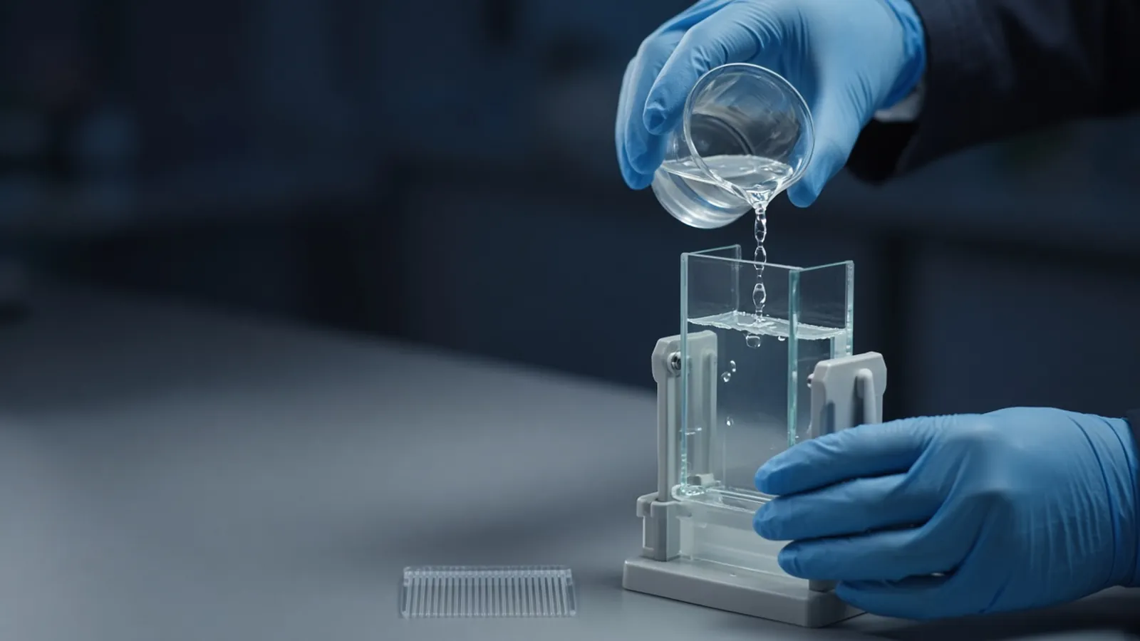
The same bacterial lysate can return three concentrations from three honest kits. Bradford, BCA and absorbance at 280 nm weigh different chemical facts, and a lysate is full of membranes, detergents, reductants and nucleic acids that each assay notices differently. This page is about which interference dominates and how to pick the assay the buffer allows. It is a research comparison. Follow the insert that came with the kit you open.
The wider measurement landscape is in how laboratories estimate protein concentration. A number you then load on a gel is interpreted in reading a protein gel. Buffer composition, which decides the interference, is recorded as in preparing a buffer and checking pH.
What each method is actually seeing
The Bradford assay uses Coomassie dye in acid. The colour shift depends strongly on basic and aromatic residues. Proteins that are rich in arginine respond more than proteins that are not, so two pure proteins at the same mass need not give the same colour. In a lysate the dye reports a residue-weighted mixture, expressed as equivalents of whatever standard you plated. Ionic detergent, SDS in particular, disturbs the dye equilibrium at concentrations typical of denaturing lysis buffers. Many inserts restrict SDS to a low level well below a 1 percent lysis. Reducing agents are usually less troublesome for Bradford than for BCA, which is why a DTT-heavy buffer sometimes forces the choice.
The BCA assay, bicinchoninic acid, detects copper reduced by peptide bonds and by some side chains, under alkaline conditions. It is often more tolerant of non-ionic and some ionic detergents than Bradford is, within limits printed on the kit. It is famously sensitive to reductants. DTT, beta-mercaptoethanol and some reducing sugars reduce copper and inflate the reading, sometimes wildly. Chelators that starve the copper chemistry push the reading the other way. A lysate in a commercial extraction buffer can contain both a helpful detergent and a hidden reductant. The detergent is not the dominant interference if the reductant is present.
Absorbance at 280 nm reports aromatic side chains, mostly tryptophan and tyrosine, plus a contribution from disulfides nearer 260 to 280. For a pure protein with a known sequence, an extinction coefficient turns that absorbance into a concentration, with assumptions spelled out in any careful methods note: the protein is folded or you accept the coefficient you calculated, the buffer blank is real, and nothing else absorbs. A crude lysate violates the last assumption. Nucleic acids absorb strongly near 260 and still contribute at 280. Membrane fragments scatter light. Triton and NP-40 absorb in the ultraviolet because of their aromatic rings. The cuvette can be "following Beer's law" and still not be measuring your protein.
Which interference dominates in a dirty lysate
Rank the buffer before you rank the proteins. If the tube contains SDS at lysis strength, Bradford is the method most likely to be compromised, and A280 may also be messy if the lysate is turbid. BCA may be the least-bad chemical assay if reductant is absent and the kit lists that SDS level as compatible. If the tube contains DTT or beta-mercaptoethanol at typical inhibitor or sample-prep levels, BCA is the method most likely to be inflated, and Bradford becomes the more defensible colour assay, provided detergent is low enough. If the tube is a clarified, detergent-free extract and you only need a rough load for a gel, either colour assay can serve, and A280 remains a mixture reading you should label as such.
Nucleic acids dominate A280 more than they dominate Bradford. A viscous, uncleared lysate should not be quantified by a direct 280 nm reading and then called milligrams of protein. Membranes dominate by scatter: a cloudy cuvette has an absorbance that is partly physics. Spin or filter before you believe a spectrophotometer, and still treat the number as provisional.
Inclusion-body washes add another matrix. Urea or guanidine, residual detergent, and a protein that is one species rather than a lysate proteome change which assay is fair. A pure inclusion-body protein in denaturant can be a better A280 sample than the lysate it came from, if the denaturant blank is subtracted and the coefficient is the one for that sequence. The lysate of the same culture is not that sample.
Why two kits disagree while everyone followed the instructions
Each kit asks you to build a standard curve and to incubate for a stated time at a stated temperature. Doing that correctly makes the curve valid for the standard protein in the standard buffer. It does not make BSA behave like your mixture, and it does not cancel a reductant the standard wells never contained. A lysate diluted into the assay so that SDS or DTT falls inside the kit's tolerance can still sit on a steep part of the interference curve. One person dilutes tenfold, another fourfold, and both "follow the kit" while the interfering component is unequally diluted.
Colour development is also timed. Bradford readings drift. BCA colour keeps developing and depends on temperature. A warm afternoon on the bench is not the room the curve was built in if the plate then sat by a window. Parallel timing of standards and samples matters more than a second decimal place.
The practical rule is small. Pick the assay the buffer allows, using the compatibility table in your insert. Run the lysis buffer alone and the lysis buffer spiked with a known mass of standard. If the spike is not recovered within an error your experiment can tolerate, change assay or change buffer. Then name the standard in every table: Bradford, BSA-equivalent, or BCA, BSA-equivalent, or A280 using a stated coefficient. Two numbers may both be real and both be the wrong input for a stoichiometric calculation.
| Matrix in the lysate | Bradford | BCA | A280 |
|---|---|---|---|
| SDS at typical lysis levels | Often incompatible; check the insert | Often more tolerant; check the insert | Scatter and buffer absorbance remain |
| DTT or beta-mercaptoethanol | Usually the more robust colour choice | Signal inflated by reductant | Little direct effect from the reductant |
| Nucleic acids | Minor compared with A280 | Minor compared with A280 | Major contribution at 280 nm |
| Triton or NP-40 | Possible dye effects; check the insert | Often tolerated within a stated limit | Strong ultraviolet absorbance |
| Clarified, dilute, no detergent | Usable as a BSA-equivalent | Usable if reductant is absent | Still a mixture, not a single protein |
Branch when the number will be used for a decision
If the number only sets a gel load, a BSA-equivalent colour assay in a compatible buffer is enough, and a twofold disagreement is a reason to load a small series rather than to average the kits. If the number sets a specific activity, the assay and the standard are part of the result. Changing from Bradford to BCA halfway through a project creates a fake jump in purity. If the number claims a yield of one recombinant protein from a lysate, remember you measured the proteome, weighted by the assay. The target may be a thin band. Pair the number with a gel.
When a spike of standard into the lysate buffer misses, stop. Either the interference dominates or the pipette and the curve are at fault. A buffer-only blank that already equals the lowest standard is an interference result. Dilute further only if the kit's linear range still contains the diluted sample. If it does not, switch assay class.
Research limits
These assays estimate polypeptide mass in research samples. They are not endotoxin tests, not activity assays, and not a release specification for a medicine. A high reading can be reductant. A low reading can be a protein the dye barely sees. Report the method. Colour reagents and copper solutions are chemical hazards under local rules.
Kits, heat, and a curve built in the morning
BCA colour depends on temperature. A curve incubated at a controlled temperature and samples left on a warm bench are not the same calibration. In a hot laboratory, follow the insert's incubation and keep the plate out of direct sun. Humidity does not change Beer's law. It does fog cuvettes taken from a cold room, and a wet optical face is a false absorbance. Wipe and blank again.
If a power cut stops a heated incubator mid-BCA, the partial colour is not the kit's endpoint. Restart the timing decision from the insert rather than reading a plate that cooled halfway.
What to send when you need reagents or a measured protein
State the buffer ingredients that constrain the assay: SDS, urea, DTT, glycerol, and the standard you intend to report. Those chemical classes live in the reagents and chemicals catalogue. If a collaborator will express the protein and you need the concentration defined a certain way, say so in the expression and purification enquiry reference, which is a place to discuss the work, and repeat the assay name in the quote request. A yield without the assay and the standard is not yet a specification.
Questions from the bench
Why can Bradford and BCA both be in range and still disagree by twofold?
They respond to different chemical features, and a lysate supplies different interferences to each. A kit curve can be perfect against its own standard while the lysate dye response is not the same as that standard. The disagreement is a property of the matrix until you show otherwise.
Is A280 ever the right reading on a crude lysate?
It is a poor abundance assay for a crude lysate because nucleic acids, scatter from membranes, and some detergents absorb or deflect light in the same window. A280 becomes more honest after the protein is purified and you have an extinction coefficient for that sequence. Use it on a lysate only as a rough, flagged estimate.
Which standard should I name in the notebook?
Name the protein in the standard tube, usually BSA or immunoglobulin, and the assay. A Bradford number against BSA is not a mass of your enzyme. If the downstream calculation needs absolute mass, say that the figure is BSA-equivalent, or calibrate with the purified protein itself.
Do protease inhibitors or glycerol break these assays?
Some inhibitor components and high glycerol change colour development or blanks. Compatibility is a table in the insert, not a universal yes. Run the buffer, including additives, as a blank and as a spiked standard before you trust the lysate number.
References
Manufacturer names identify published method classes. Trademarks remain with their owners. Catalogue records on this site are independent references for enquiry. They are not a statement of inventory, distribution rights or a supply commitment. This page is educational. It is not medical advice, a diagnostic protocol or a biosafety approval.
Catalogue
Related products and categories
These links follow the subject of the article into published manufacturer references. A listing is a reference for an enquiry, not a statement of stock or distribution rights.
Continue in this cluster
Related reading
Reading a protein gelHow to read a protein gel: what SDS-PAGE bands, smears, ladders and loading differences can support, and what they cannot identify. How laboratories estimate protein concentrationHow laboratories estimate protein concentration with absorbance, dye-binding and copper assays, and why the number depends on the standard.
 Preparing a buffer and checking pHPrepare a buffer at a stated pH and temperature, record the salt form, and treat a drifting electrode or a bad slope as a failed control.