
selection guide
Fluorometric versus absorbance quantification
Choose a dye assay when double-stranded DNA must be counted, and absorbance when you need a fast check of everything that absorbs at 260 nm.
- Author
- EVRINTH Editorial Team
- Published
- 8 October 2026
- Updated
- 8 October 2026
- Reading time
- 8 min
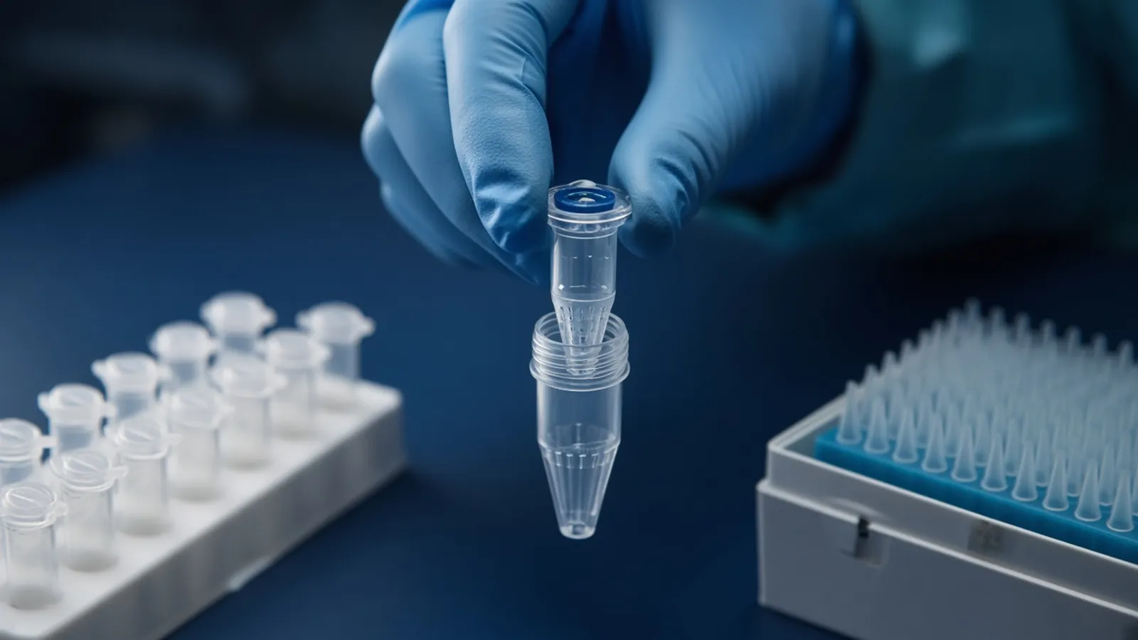
Fluorometric versus absorbance quantification is a choice about what you are willing to count. A dye assay with a standard curve reports double-stranded DNA more selectively and misses most free nucleotides. Absorbance is fast and counts everything that absorbs at 260 nm, including phenol and nucleotides. Pick the one the downstream assay actually needs. When the two numbers disagree, the sample is telling you it is dirty, and that disagreement is data. This is a research selection guide, not a kit insert and not a clinical measurement.
Nucleic acid extraction sets up the disagreement. A silica column that was overloaded or poorly washed hands you a tube full of chaotrope, short fragments, and the DNA you wanted. The method classes are laid out in how DNA extraction methods differ. Quantification does not repair that tube. It tells you which claim you can make about it.
What a dye is counting
Fluorometric dyes used for double-stranded DNA bind that structure and fluoresce much more once bound. You excite the sample, read the emission, and compare it with a standard curve built from a known double-stranded standard in the same buffer. Without the curve, you have a brightness. With the curve, you have a concentration inside the range the standards cover. Samples above the top standard need dilution. Samples below the bottom standard are not rescued by drawing the line through zero and hoping.
Selectivity is the point. Free nucleotides and very short pieces that do not hold the dye contribute little. RNA and single-stranded DNA contribute less than they would to an absorbance reading, to a degree that depends on the dye class. Follow the reagent note for the cross-reactivity it actually publishes. Do not assume every fluorescent DNA dye ignores RNA equally.
The standard has to match the question. A plasmid standard and a genomic standard of the same nominal mass can still sit differently in some assays if the dye's preference for length is strong. Record which standard you opened. A curve made last week, in another buffer, is a different experiment.
What absorbance is counting
An absorbance reading at 260 nm is a bulk optical sum. Double-stranded DNA, single-stranded DNA, RNA, primers, and free nucleotides all contribute. Phenol, which absorbs nearer 270 nm, leaks into a 260 reading when enough of it remains. The conversion factors people use for a 1 cm path — about 50 ng per microlitre per absorbance unit for double-stranded DNA, about 40 for RNA, about 33 for single-stranded DNA — assume the absorbance is mostly the molecule named in the factor. The pathlength setting has to match, or the factor is being applied to the wrong absorbance.
The speed is real. There is no curve to build and no dye to protect from light. Ratios come from the same spectrum. A260/A280 near 1.8 for DNA and near 2.0 for RNA hints at protein. A260/A230 often falls when chaotrope, phenol, or carbohydrate remains. Those ratios are cleanliness hints. They are not RNA integrity, not fragment length, and not proof that a polymerase will work.
Absorbance is the right screen when you need a fast check, you have a few microlitres to spare, and a later step can tolerate some non-DNA absorbance. It is a weak way to dose a library or a ligation when the extraction was dirty.
Choose by the assay that consumes the tube
Write the downstream method before you pick the reader.
A hardy endpoint PCR often needs only that some template is present. Absorbance, or even a dilution series, may be enough. A dye number will not tell you whether the target sequence is there.
A restriction digest or a ligation needs a mass of the DNA the enzyme will see. If ratios are already in the familiar window and the gel shows one species, absorbance is often a fair dose. If ratios are off, or the prep was a plasmid alkaline lysis with leftover RNA, a double-stranded dye is the fairer mass. RNA integrity does not belong in that plasmid sentence. RNA in the tube is a contaminant that absorbance will count and a double-stranded dye will mostly skip.
A short-read or long-read library usually specifies double-stranded input. Dose it with a selective dye, then judge length on a trace or a gel. An absorbance dose that includes adapter dimers and free nucleotides will under-load the true library and overstate how much you added.
An RNA assay needs an RNA-selective dye or an absorbance factor of about 40, plus a separate integrity check. A double-stranded DNA dye will miss most of the RNA and send you into a library with too little material. A high RNA mass still says nothing about whether the ribosomal peaks survived.
| Downstream need | Prefer | Why the other number misleads |
|---|---|---|
| Fast cleanliness screen | Absorbance and the two ratios | A dye will not show chaotrope at 230 nm |
| Double-stranded mass for a library or a ligation | Dye plus a same-day standard curve | Absorbance counts nucleotides, RNA, and phenol |
| Target copy number | The amplification assay itself | Both bulk methods count the whole tube |
| RNA mass | RNA dye or the RNA absorbance factor | A DNA dye ignores most RNA |
| Fragment length or RNA integrity | Gel, trace, or an electrophoretic score | Neither optical method measures length |
When the two numbers disagree
Read the direction of the gap.
Absorbance high, dye low: something in the tube absorbs at 260 nm and is not double-stranded DNA the dye recognises. Free nucleotides after a DNase step, residual RNA, or phenol are the usual suspects. Trust the dye for double-stranded mass, and treat the gap as a purity finding. A silica column wash that was skipped often looks exactly like this.
Dye high, absorbance low: check the curve, the dilution arithmetic, and whether the absorbance pathlength was applied twice or not at all. A very clean sample can also sit below the range where a short-path pedestal is steady, while the dye curve still covers it. Do not average the two numbers into a compromise that matches neither method.
Both low: there is little of what either method sees. That can be a failed extraction, a huge elution volume, or a sample that was never there. Both high and ratios clean: you may simply have a rich prep. Confirm with a dilution that lands inside both ranges before you celebrate.
The disagreement is not a reason to pick the figure that makes the next protocol happy. Record both, name the method you will dose from, and keep the other as the caution.
A short workflow
Measure the blank or the dye background in the elution buffer, not in a random water. Build the dye curve before the samples, with at least the low and high standards the reagent note requires. Read absorbance on a clean pedestal or a stated cuvette path, and write down the factor.
If the assay is a library or a ligation, dose from the dye when the two methods disagree. If the assay is a purity screen before you decide whether to clean the tube, keep the absorbance spectrum. If you need both claims, run both and file them as separate results. Do not subtract one from the other and call the remainder a concentration.
Branch when the dye curve is flat or the standards miss their expected window. The lamp, the dye, or a degraded standard is the problem. Sample numbers from a failed curve are not low yields. Make a fresh curve. On the absorbance side, branch when the blank looks like nucleic acid. Clean the surface and reblank before any sample is believed.
Failures that look like biology
A dye left in the light, or a standard thawed repeatedly, compresses the curve so every sample shifts together. That pattern is the assay, not every extraction. Too much DNA also plateaus a dye, and a rich prep looks moderate until you dilute into the standards. A short-path absorbance reader is noisy on a very dilute eluate. Phenol and chaotrope can leave a dye mass looking fine while the next polymerase still dies, so keep the 230 nm shoulder or a small enzyme test in the decision.
Safety and research limits
Dyes that bind nucleic acids are chemical reagents with their own safety data. Treat the tube you have, not a generic "DNA stain" story. Intercalating dyes are not automatically safe because the volume is small. Absorbance readers keep their ultraviolet lamp enclosed. Do not bypass the housing.
Specimens stay under the biosafety decision your institution already made. A quantification method does not inactivate a pathogen and does not make a research number into a diagnostic quantitation. The WHO laboratory biosafety manual is background for the institutional conversation, not permission for this bench.
Heat and a curve that drifted
A curve made in a hot room with a warm pipette is a calibration error shared by every sample. Bring the dye to the temperature the note states. If the power fails mid-read, measure the curve and the samples again in one session.
What the enquiry should name
Say whether you need a double-stranded dye kit, an RNA dye, or only absorbance consumables, and name the downstream method and the elution buffer. State the expected mass range so the standard window is part of the request. Browse the sample preparation catalogue, connect the prep to the assay on the sample preparation pathway, and send the detail with the quote request. The nucleic acid isolation enquiry reference can carry the specimen and the assay together. It is an enquiry reference. Ask whether a quotation is possible.
Questions from the bench
Why is my absorbance number higher than the dye assay?
Absorbance at 260 nm counts double-stranded DNA and also free nucleotides, RNA, and some leftovers such as phenol. A double-stranded dye ignores most of those. The gap is evidence that the tube is not pure long DNA. Clean the sample or choose the number that matches the assay you will actually run.
Can I skip the standard curve if I only need a rough figure?
A fluorescence reading without standards is a relative brightness, not a concentration. The curve is what turns that brightness into mass, and it has to be in the same buffer and on the same day as the samples. If you need only a presence check, absorbance is the faster tool and should be named as such.
Which number should I use for a sequencing library?
Library kits usually ask for double-stranded DNA mass. A dye selective for that molecule is the fairer input, because leftover primers and nucleotides would inflate an absorbance figure. Confirm fragment length on a separate trace. Neither method proves the inserts are the sequences you intended.
Does a dye assay replace an RNA integrity score?
No. A dye can estimate RNA mass when you chose an RNA-selective reagent and a matching standard. Integrity, including an electrophoretic score, asks whether the molecules are still long. A bright RNA signal can come from fragments that will not build the library you planned.
References
Manufacturer names identify published method classes. Trademarks remain with their owners. Catalogue records on this site are independent references for enquiry. They are not a statement of inventory, distribution rights or a supply commitment. This page is educational. It is not medical advice, a diagnostic protocol or a biosafety approval.
Catalogue
Related products and categories
These links follow the subject of the article into published manufacturer references. A listing is a reference for an enquiry, not a statement of stock or distribution rights.
Continue in this cluster
Related reading
How DNA extraction methods differHow silica columns, beads, precipitation and organic extraction change DNA yield, length and inhibitors, and which class fits the assay you actually plan toA glossary of purification termsDefine chaotrope, bind-wash-elute, yield, purity ratios and RIN, then walk a purification so those words describe one real workflow.Blood saliva and tissue as starting materialExplain how EDTA blood, heparin blood, saliva and solid tissue behave as extraction inputs, and why a serum tube is not whole blood.