
Pillar guide
How DNA extraction methods differ
How silica columns, beads, precipitation and organic extraction change DNA yield, length and inhibitors, and which class fits the assay you actually plan to
- Author
- EVRINTH Editorial Team
- Published
- 8 October 2026
- Updated
- 8 October 2026
- Reading time
- 8 min
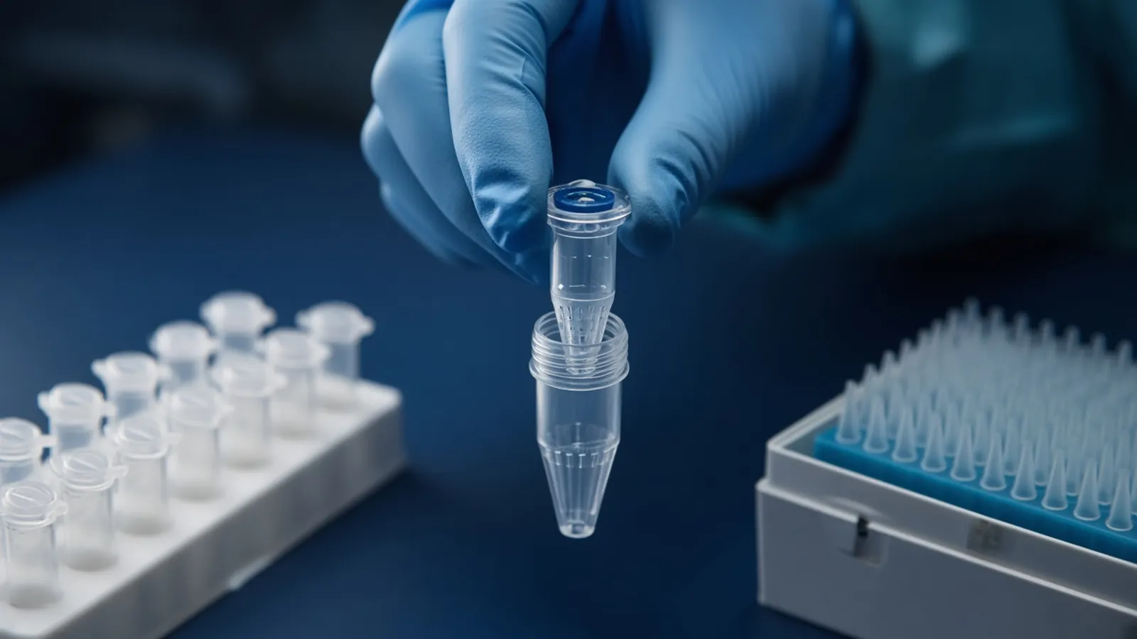
DNA extraction is the decision about what you are willing to lose. Every method lyses a specimen, then separates nucleic acid from protein, lipid and the small molecules that would poison the next enzyme. Columns, beads, alcohol precipitation and organic solvents do that separation differently, so they return different yields, different fragment lengths and different leftovers. This page is a way to choose among those classes. It is a research explainer, not a kit insert and not a clinical sample protocol.
The downstream assay picks the winner. A short PCR can tolerate sheared DNA and still fail on a trace of ethanol. A long-read library can forgive a lower yield and still fail if the fragments were broken in the lysis. RNA co-purified by accident is a separate problem, taken up in protecting RNA during extraction.
Sourcing starts from the sample preparation catalogue. Send the specimen and the assay with the quote request. A catalogue family name is not a promise about a single formulation.
What every method is trying to do
Lysis has to open the protein and membrane around the DNA. Detergent, chaotrope, alkali, enzyme, heat or grinding each open a different matrix. Plant tissue needs a way past cell walls and polyphenols. Bacteria with tough walls need an enzyme or a mechanical step that mammalian cells may not. A buffer that works on a cultured cell pellet can leave a tissue chunk looking extracted when most of the DNA is still inside.
Once the DNA is free, the method has to keep it and discard the rest. Silica chemistry binds DNA in high chaotropic salt and releases it in low salt. Magnetic beads use a related bind-and-wash idea, or a crowding-agent chemistry that can also select by size. Alcohol precipitation can preserve long molecules and can also carry polysaccharides. Organic extraction partitions proteins into a phenol phase and leaves nucleic acid in the aqueous phase only if the pH matches that protocol.
Each method has a leftover it is bad at removing. Knowing that leftover is more useful than memorising a brand name.
How the classes differ in practice
A silica spin column is fast and has a finite binding capacity. Too much lysate, and DNA passes through while you record a low yield. The wash that removes chaotrope usually contains ethanol, and ethanol left in the eluate inhibits polymerases and ligases. A dry spin helps. Over-drying the membrane can make elution poor in the other direction. Elute in the buffer the next assay accepts. Tris-EDTA protects stored DNA and can also chelate magnesium the next enzyme needs, so storage buffer and assay buffer are allowed to differ.
Bead methods share that wash logic. A magnet replaces the column frit, which suits automation when the magnet and the tube match. Silica-paramagnetic beads behave like a column. Carboxyl beads used with a crowding agent and salt bind in a size-biased way: more crowding agent pulls in shorter fragments. That bias helps when you are removing primer-dimer and hurts when you wanted those short pieces. Beads do not repair a bad lysis.
Precipitation methods, including salting-out followed by alcohol, remain rational when you want long genomic DNA and can accept a pellet that is slow to dissolve. The pellet can be invisible, or mostly salt. A careful wash and patience during resuspension matter more than a vortex that shears the DNA you meant to keep long. Residual salt and ethanol are the usual reasons the next enzyme is quiet.
Organic extraction with phenol and chloroform is effective and hazardous. The aqueous layer holds DNA only under the pH of a DNA protocol. Acidic phenol recipes used for RNA move DNA the other way. The interphase holds protein, and pipetting through it carries contamination. Only laboratories that already have the cabinet, the waste path and the training should treat this as a default.
Crude lysates, including hot alkaline lysates and chelating-resin preps, can feed a hardy PCR and still be the wrong tool for quantity, cloning or a scarce sequencing target. Inhibitors travel with the crude material. A dilution is often the real purification step.
| Method class | What it tends to protect | What it tends to leave behind or break |
|---|---|---|
| Silica column | Speed, small elution volume, removal of many proteins | Residual ethanol, sheared DNA if lysis was harsh, overload losses |
| Magnetic beads | The same bind-and-wash logic, easier scaling | Size bias on crowding-agent beads, carry-over if washes are skipped |
| Alcohol precipitation | Long fragments, simple reagents | Salt, ethanol, polysaccharides, a pellet that is slow to dissolve |
| Phenol-chloroform | Difficult matrices when the lab can handle the solvent | Phenol carry-over, interface contamination, chemical hazard |
| Crude lysate | A quick PCR from a robust target | Inhibitors, poor storage, no claim about purity |
A workflow with branch points
Start from the specimen, not from the kit that happens to be in the freezer. Record input mass or cell number, because "low yield" is meaningless without it. Lyse until the matrix is actually dispersed. A clear lysate from tissue that still has a core is not a complete lysis.
If the yield is low and the input was high, suspect overload or incomplete lysis before you suspect the elution buffer. Split the lysate or repeat the lysis on a matched piece and see which change moves the number. If the input was tiny, you may simply be under the practical range of that chemistry.
If absorbance looks high and PCR fails, suspect inhibition. Dilute the eluate tenfold and try again. A recovery after dilution means inhibitors or too much DNA, not an absent target. A blank extraction — water or buffer taken through every wash — belongs in the same PCR. A band from the blank is contamination acquired during the prep, and the sample bands are not interpretable until that is solved. The logic is the same as in PCR controls and contamination control.
If the DNA must stay long, stop vortexing genomic preps and judge length on a gel or a fragment trace. A 260/280 ratio near the familiar DNA window is a hint about protein, not a length measurement and not an inhibitor test. RNA in the same tube inflates the 260 reading.
Failure modes worth naming
Brown or viscous plant eluates often still contain polyphenols or polysaccharides that bind DNA and the next polymerase. Use a plant-aimed method or a cleanup chosen for that contaminant.
A smear of very short DNA on a gel means the specimen was already dead and degraded, or the lysis was violent, or a nuclease stayed active because the chaotrope or the heat step never fully stopped it. Preserving specimen condition before lysis beats any column. Tissue left warm on a bench has already decided the fragment length.
If a downstream enzyme dies after an organic prep, assume solvent carry-over until a cleanup says otherwise. Guanidinium left by a skipped wash does the same quiet damage and often shows up as a poor 260/230 ratio. That ratio is another hint, not a verdict. Plasmid alkaline lysis is a different procedure from genomic extraction. Name the molecule you want before you name the kit.
Safety, climate and the enquiry
Chaotropic salts are harmful. Phenol and chloroform are worse. Follow the hazard note on the bottle you opened, and follow institutional waste rules. Specimens from people or infected animals stay inside the biosafety decision your committee already made. The WHO laboratory biosafety manual is a public reference for that institutional conversation. This page does not approve a containment level.
Washes dry slowly in humid air, so a column that looks dry can still hold ethanol, and the next polymerase stalls. A centrifuge that stops mid-run because the power dropped has not finished the separation. Restart only if you know which fraction is which.
EVRINTH can take a sourcing question. State the specimen, the input range, the fragment length you need, the downstream assay, and any inhibitor you already know is in the matrix. The nucleic acid isolation enquiry reference is a place to frame that question. It is an independent method reference. Ask whether a quotation is possible. Do not read it as a statement that a preparation service is already running. The sample preparation pathway connects extraction to the assays that consume the eluate.
Choose a DNA extraction class for the next assay
- 01Name the downstream question firstWrite whether you need PCR, a clone, a long intact fragment or a sequencing library. Yield, fragment length and inhibitor tolerance are different goals and they pick different methods.
- 02Match lysis to the specimenCells, tissue, plants, blood and bacteria do not break open the same way. Choose a lysis class that opens that matrix without assuming one buffer fits all of them.
- 03Pick the separation chemistryUse silica or a bead bind-and-wash when you need a short, inhibitor-poor DNA. Use a gentler precipitation route when length matters more than speed. Treat organic extraction as a hazard decision as well as a chemistry decision.
- 04Define the failure check before you eluteDecide what you will measure: a blank extraction, a gel or trace for length, and a small downstream test. A high absorbance number alone is not a pass.
Questions from the bench
Does a high A260 reading mean the DNA will amplify?
No. Absorbance at 260 nanometres estimates nucleic acid and also responds to free nucleotides and some contaminants. Inhibitors such as ethanol, heme and polysaccharides can leave the number looking fine while the enzyme in the next tube does nothing. A blank extraction and a small PCR or digest tell you more.
Why would two silica kits give different yields from the same blood tube?
Binding chemistry, wash stringency, lysis time and the input window all differ between kits that share a family name. Overloading a column past its binding capacity looks like a low yield even though the sample was rich. Compare methods on a split specimen, and record the input amount you actually used.
When is organic extraction still a rational choice?
It remains useful for some difficult tissues and for preparations where a laboratory already has the hood, the waste stream and the skill to handle phenol safely. It is a poor default when a column or bead method already meets the fragment length and purity the assay needs. The hazard is part of the method, not a footnote.
Can the same prep be used for RNA-seq if the gel looks intact?
DNA integrity is not RNA integrity. A genomic smear that looks acceptable says little about whether RNases destroyed the transcripts in that tube. RNA needs its own handling, described in the companion note on protecting RNA during extraction.
References
Manufacturer names identify published method classes. Trademarks remain with their owners. Catalogue records on this site are independent references for enquiry. They are not a statement of inventory, distribution rights or a supply commitment. This page is educational. It is not medical advice, a diagnostic protocol or a biosafety approval.
Catalogue
Related products and categories
These links follow the subject of the article into published manufacturer references. A listing is a reference for an enquiry, not a statement of stock or distribution rights.
Continue in this cluster
Related reading
 Protecting RNA during extractionHow to keep RNA intact from lysis to elution: RNase control, what A260 ratios do not prove, and the checks that show whether the preparation is still usable.A glossary of purification termsDefine chaotrope, bind-wash-elute, yield, purity ratios and RIN, then walk a purification so those words describe one real workflow.Blood saliva and tissue as starting materialExplain how EDTA blood, heparin blood, saliva and solid tissue behave as extraction inputs, and why a serum tube is not whole blood.