
troubleshooting
Fraction collection and how to assay fractions
Decide whether a fraction is worth pooling: collect by volume or peak, then assay activity, a gel, or absorbance before you combine.
- Author
- EVRINTH Editorial Team
- Published
- 8 October 2026
- Updated
- 8 October 2026
- Reading time
- 7 min
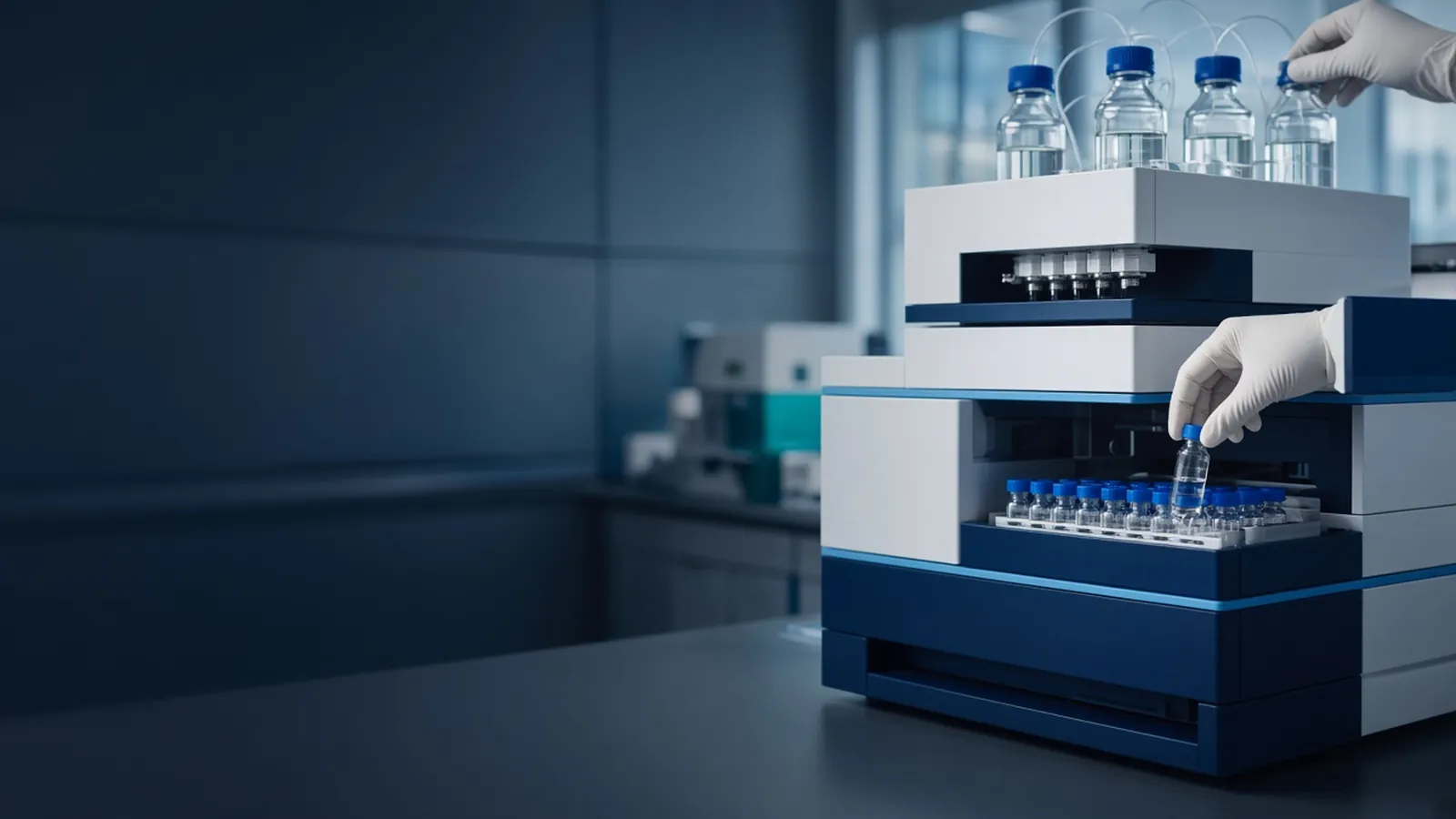
A fraction is a tube of mobile phase that left the column during a defined slice of the run. The chromatogram suggests which tubes to look at. The assay decides which tubes to keep. This page is the troubleshooting path when those two disagree. The separation that filled the tubes is described in chromatography methods in life-science labs.
Two ways to cut, and the delay between them
You can advance the collector every fixed volume, or every time the detector says a peak started and stopped. Fixed volume misses nothing that elutes, including activity that has no chromophore, and it spends tubes on baseline. Peak triggering saves tubes and will throw away a colourless active protein if the threshold never fires. For a new method, collect by volume across the window where binding and elution should occur, assay, and only then trust a peak trigger.
The detector is upstream of the drip. The volume of tubing between the flow cell and the tube is a delay. At analytical HPLC flows that delay can move the peak by a large part of a narrow fraction. At preparative flows it is a smaller fraction of a big tube and still enough to put the front of a peak into the previous tube. Measure it. A dyed step or a salt step you can see, collected by volume, shows which tube actually received the change. Write that offset into the collector programme. ISO 8655-1 is relevant when a pipette, not the collector, is what you later use to build the assay plate. The collector's own calibration is a separate record.
Column volume keeps the slice honest. A fraction of 0.05 column volumes is a fine analytical cut. A fraction of half a column volume on a polishing column blends the separation you just paid for. Match tube size to peak width. A peak that is three tubes wide can be assayed tube by tube. A peak that is one-tenth of a tube cannot be rescued by a clever threshold.
What to assay, and in which order
Absorbance is the fastest location tool. At 280 nanometres it sees aromatic protein and also sees imidazole and other additives. At peptide-bond wavelengths it sees almost every peptide and a great deal of buffer noise. Use it to choose which tubes are worth a slower assay. Do not use it as the only reason to pool.
A gel asks whether the band you expect is in the tube and what else is with it. Load equal volumes, or equal protein, and say which you did. A thick band from a large volume can be a dilute contaminant. Activity asks whether the function survived. It is the right decider for an enzyme and a poor decider for a structural protein that has no assay yet. A blot asks about an epitope. Mass spectrometry asks about mass and, with fragments, about identity, which is a claim you report with the care HUPO discusses, against a sequence you can check in UniProt.
Run the assay on the load, the flow-through, a baseline tube, the leading edge, the apex, and the trailing edge. That set distinguishes "never bound", "still on the column", "in the shoulder", and "the assay does not work in this buffer". Spiking a known amount of load into a baseline fraction tests inhibition. If the spike dies, the elution salt or the imidazole is the story, and desalting one tube is the next check. Chromatography delivered the liquid. It did not promise the assay would tolerate the liquid.
Symptoms and the next check
Activity only in the flow-through means the column did not bind the target. Stop polishing the elution tubes. Fix pH, salt, solvent, or the tag, and rerun at scout scale. Activity only on the trailing edge, with the ultraviolet apex earlier, means the detector and the function are not the same molecule, or the delay volume put the protein one tube later than the trace. Shift the assignment by the measured delay and reassay before you change the gradient.
Activity split across two regions can be two forms, an aggregate and a monomer, or a collection glitch that skipped a tube. Look at the gel of both regions before you call them isoforms. A gap in tube volumes, or a tube that overflowed into its neighbour, is a hardware symptom. Fix the rack and the flow before you invent a biological story.
Empty tubes under a beautiful HPLC peak usually mean the collector never saw the trigger, the diverter valve stayed in waste, or the peak was so narrow that the delay put it between cuts. Watch one run with the lid open if that is safe for the solvent. A peak in the waste line is a valve problem. A peak in a tube you did not expect is a delay problem.
Pooling too early is the failure that looks like productivity. You combine ten tubes because the trace was above threshold, and the gel of the pool grows a contaminant that was only in tubes eight and nine. The fix is to keep the individuals until the assay sheet says they agree. A pool cannot be unmixed.
| What you hold | Question it can answer | Question it cannot |
|---|---|---|
| Absorbance trace | Where chromophores left the column | Which tube has activity, or whether co-elution happened |
| Equal-volume gel | Which tubes contain the band, and the neighbours | A quantity, unless you calibrated the stain |
| Activity with a spike control | Where function is, and whether buffer kills it | Identity of a band that is not the enzyme |
| Mass result on one tube | A mass consistent with the sequence | That neighbouring tubes are the same species |
| A pool made first | Almost nothing reliable | Purity, yield, or which cut was the mistake |
Cold tubes and a warm assay
Fractions warm up, evaporate, and grow if they sit in open racks. Cap them, hold them at the temperature the protein prefers, and assay before you leave them over a weekend. A collector in a hot laboratory will concentrate late tubes by evaporation and make the last fraction look richer than it is. Weigh or read the volume of a suspicious tube. Organic fractions lose volume faster than aqueous ones and may belong in a hood, not on an open bench.
A power cut that stops the pump and the collector at different moments creates a tube of unknown volume. Mark that tube and do not average it into the pool. Restart from a saved sample only if you still have one. The half-collected gradient is not a calibration.
Research-use limits
Assays on fractions support a research keep-or-discard decision. They are not a clinical result. The biological risk of the fraction is the risk of the load, until your institutional rules say it has changed. Chemical risk follows the mobile phase. Do not dry a solvent fraction on the bench because the assay volume was small.
Enquiry
State flow, fraction volume, detector-to-tube delay if you know it, tube material, and whether collection is by volume or by peak. Name the assay you will run so the fraction buffer can be discussed against it. Hardware is in the scientific instruments catalogue. Send the requirement with the quote request. If the fractions are a protein purification record, the custom protein expression and purification reference is a place to frame the method question as a discussion. ---
Questions from the bench
Should tubes be advanced by time, by volume, or by the peak?
Volume is the fair cut when flow is steady, because each tube then holds the same slice of the bed's output. Time matches volume only while the pump delivers the set flow. Peak-based collection drops tubes where the detector crosses a threshold, which is right when you trust the detector and wrong when the signal is imidazole, a solvent step, or a bubble. If pressure was unstable, believe collected volume over the clock.
The ultraviolet peak is large and the activity assay is flat. Which is wrong?
They can both be honest. Absorbance reports chromophores, including buffer additives. Activity reports the function, which may sit in a shoulder the absorbance plot calls minor, or may have been killed by the elution buffer. Assay the load, the flow-through, and both edges of the peak before you discard the run. A dead assay in every tube including a spiked control means the fraction buffer inhibits the assay, not that the column ate the protein.
When is it reasonable to pool tubes?
Pool the consecutive tubes the assay says are the same decision: enough activity or the right gel band, and an impurity you refuse still below your line. Leave out a tube that fails that test even if it sits under the apex. Pooling by the software's integration limits imports every co-eluting neighbour inside those limits. The pool is a new sample. Label it with the tube numbers and the assay, not only with the retention time.
What should a fraction-collector enquiry include?
State flow rate, tube volume, whether you need volume cuts or detector-triggered cuts, the delay between detector and drip, and the smallest peak you must not miss. Ask for the collector's volume accuracy and chemical compatibility. Send it with the quote request. A detector without a stated delay is not a collection method.
References
Manufacturer names identify published method classes. Trademarks remain with their owners. Catalogue records on this site are independent references for enquiry. They are not a statement of inventory, distribution rights or a supply commitment. This page is educational. It is not medical advice, a diagnostic protocol or a biosafety approval.
Catalogue
Related products and categories
These links follow the subject of the article into published manufacturer references. A listing is a reference for an enquiry, not a statement of stock or distribution rights.
Continue in this cluster
Related reading
Chromatography methods in life-science labsHow life-science chromatography separates molecules on a stationary phase, and what an HPLC or column peak does not prove about purity.A glossary of chromatography termsUse chromatography terms with their real limits: retention, resolution, void, and a peak that is only a detector response. Affinity and ion-exchange protein purificationHow affinity capture and ion exchange pull a protein from a lysate, and why each collected fraction still needs an identity check.