
glossary
Plate readers absorbance and fluorescence
Absorbance, fluorescence and luminescence are different reads. A wrong wavelength or a clear plate in a fluorescence assay looks like a failed ELISA.
- Author
- EVRINTH Editorial Team
- Published
- 8 October 2026
- Updated
- 8 October 2026
- Reading time
- 9 min
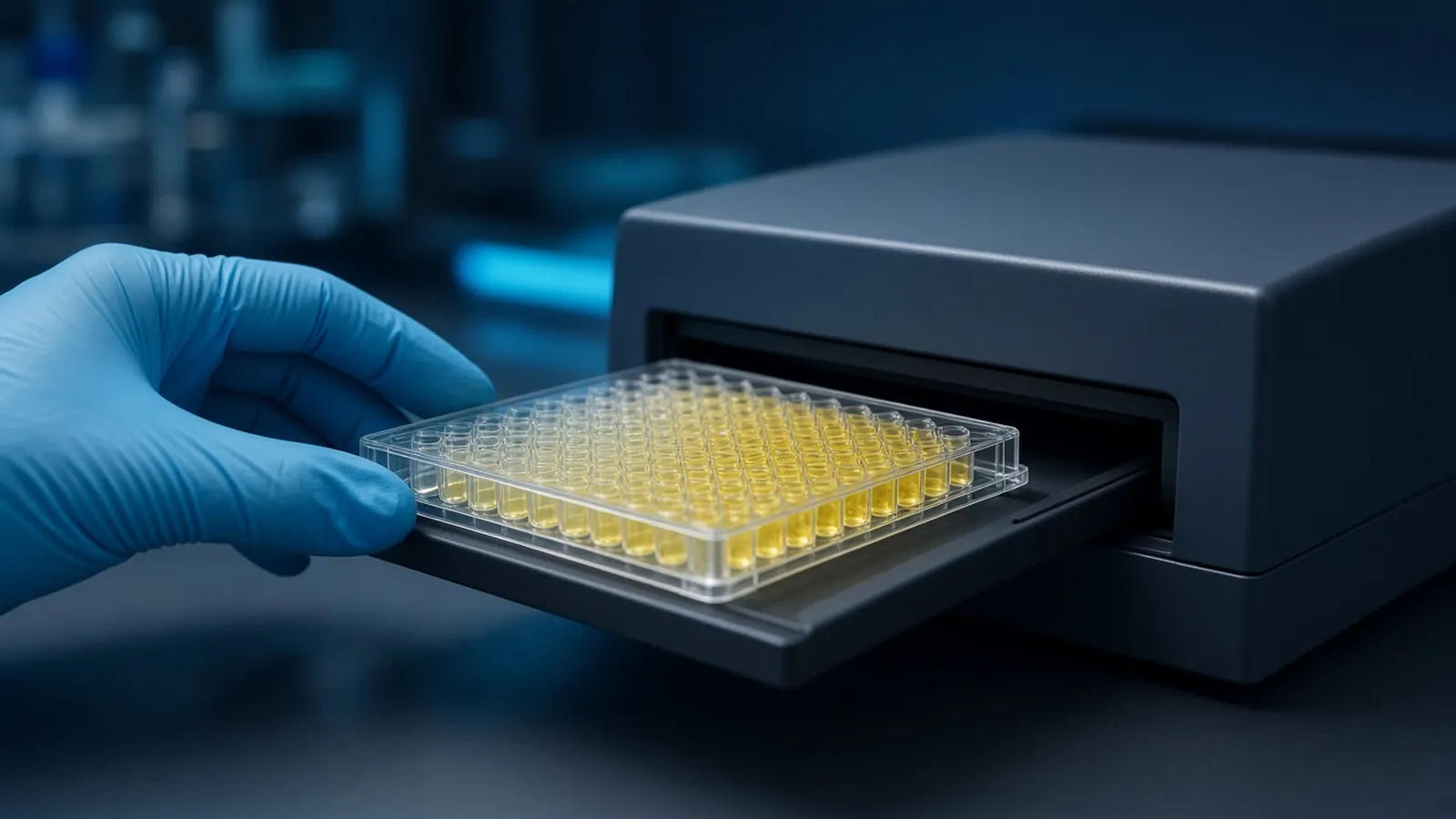
Plate readers, absorbance and fluorescence are the last translation in an immunoassay, and they are easy to misname. The drawer in the photograph closes on a plate. The instrument then does whatever programme the previous person left loaded. Absorbance, fluorescence and chemiluminescence are three different optical jobs. A glossary is only useful here if it changes a decision: which wavelength, which filters, which plate colour, and which file you are willing to call a result. The chemistry that is supposed to create the colour or the light is in ELISA formats, controls and readout. Volume errors that change path length are kin to accurate micropipetting. This page does not rank readers by sensitivity. A ranking without your dye, your plate and your blank would be advertising.
Absorbance, path length and optical density
Absorbance is a comparison between light sent at a well and light that gets through. Optical density is the name most ELISA tables use for that comparison. The well is not a cuvette with a fixed 1 centimetre path. The path length is the depth of the liquid the beam crosses, which depends on volume, meniscus and how flat the plate sits. Two wells with the same concentration and different volumes do not owe you the same optical density. Topping up, evaporating, or leaving a bubble in the beam changes the number without a change in analyte.
The primary antibody does not absorb at the assay wavelength in any way you should interpret. The coloured product does. Nonspecific binding matters because it leaves enzyme in wells that should be quiet, and that enzyme makes the same coloured product. The reader cannot tell a specific well from a sticky one. Your blank does that work. If you blank the instrument on air and also subtract a reagent blank later, write both steps. A double correction you cannot describe is not a method.
Air bubbles and a scratched lid throw the beam. Wipe the underside of the plate. Condensation from a cold plate in a humid room scatters light and imitates a high, noisy optical density. Let the plate reach the read temperature the protocol allows, then read.
Wavelength is a choice, not a default
The lamp and the monochromator or filter pick a narrow band of wavelengths. The dye absorbs some of those bands and ignores others. Reading away from the dye's peak makes a real assay look weak or blank. Set the wavelength from the substrate sheet for the chemical state in the well, stopped or still developing.
Tetramethylbenzidine, usually called TMB, is the common example for horseradish peroxidase. Before an acid stop, the product is blue and is often read in the red region, at a wavelength the sheet names. After the acid stop many sheets specify, the well looks yellow because the product now absorbs in the blue region of the spectrum. A common setting is near 450 nanometres. Follow the sheet you opened. If the sheet also asks for a reference wavelength to subtract, record that too. Omitting a required reference, or inventing one, changes the numbers. Do not copy 450 nanometres onto an alkaline phosphatase substrate that is meant to be read elsewhere. The colour you see is a hint. The sheet is the setting.
A shared reader that wakes on the last user's wavelength will silently measure your yellow plate at a fluorescence setting or at 600 nanometres. Put the wavelength in the protocol and in the file name. Look at one well by eye before you trust a flat file. If the well is yellow and the file is empty, you have an optics problem, not a missing analyte.
Fluorescence needs two wavelengths and a darker plate
Fluorescence is emission, not a shadow. The reader excites the fluorophore at one wavelength and collects a longer emission wavelength through a filter or a second monochromator. Both have to match the dye. An excitation filter for one fluorophore on a conjugate labelled with another is a blank plate with a healthy antibody. Write the pair down: excitation and emission, in that order, from the conjugate sheet.
The plate colour is part of the optics. Clear wells let light bounce into neighbours, which is crosstalk. A bright standard beside a zero can make the zero look occupied. Black plates reduce that path and are the usual choice for fluorescence. White plates are often chosen for luminescence because they reflect photons toward the detector, and they are a poor default for fluorescence for the same reason: they move light around. If you read fluorescence in a clear plate because it was what the coating protocol used, say so, and do not treat adjacent wells as independent. Redesign the map so bright and blank wells are not neighbours, or move to a plate colour the read requires. Transferring liquid to a black plate after development is a second method, with a volume step that can add error. Record it if you do it.
Gain and integration time stretch the numbers. They are not concentrations. A gain set so the top well saturates the detector flattens the curve just as too much substrate does. Set gain on a well you expect to be bright, as the instrument manual describes, and keep it constant across plates you will compare. Auto-gain that changes every plate makes the files incomparable.
Chemiluminescence is the third word
A chemiluminescent substrate emits light because of the enzyme reaction. There is no excitation lamp to tune. The reader counts photons for a set time. The signal often changes while you count, so the order of wells and the delay after substrate addition are part of the method. A kinetic glow and a flash are different shapes. Follow the substrate class. Do not put a chemiluminescent plate through an absorbance programme and interpret the tiny numbers as optical density of a colour that is not there.
Luminescence is easily contaminated by a bright room or by a phosphorescent lid. Close the drawer. Do not compare a plate read immediately with a plate that sat while you found the software dongle, unless the sheet says the glow is stable over that wait.
Promega protocols include enzyme substrates across these readout classes. Use them as examples of how a sheet pairs enzyme, substrate and read mode. protocols.io is where you can see full plate maps that name the mode. Neither one is permission to ignore the instrument in front of you.
A decision table for the read
| Readout class | What you must set | Plate colour that usually fits | Typical silent failure |
|---|---|---|---|
| Absorbance | Wavelength, and a reference wavelength if the sheet asks | Clear bottom in the beam | Wrong wavelength, short path from low volume, bubble |
| Fluorescence | Excitation and emission pair, gain held constant | Black wells to limit crosstalk | Clear plate crosstalk, wrong filter, saturated gain |
| Chemiluminescence | Integration time, no excitation | White wells often, if the sheet agrees | Absorbance programme used by habit, delay before reading |
When the file and the well disagree
Trust the well you can see when it disagrees with a careless programme, then fix the programme and reread if the chemistry is still within the time the sheet allows. A stopped absorbance plate is more patient than a glow plate. Do not average a plate read at the wrong wavelength with last week's correct file. Delete nothing. Mark the file as mis-set and repeat the read or the plate.
Crosstalk and edge evaporation can look alike, because both make position-dependent numbers. Separate them. A fluorescence crosstalk pattern follows the bright neighbours. An evaporation pattern follows the rim even when neighbours are dim. The edge-effect discussion belongs to plate handling. The reader discussion belongs to light. If both are present, fix handling and optics before you interpret the primary antibody.
Detector linearity has a top end. Numbers that pile up at the same high value are saturated measurements. Dilute the sample or shorten the development. Do not claim those wells are equal biology.
Public measurement laboratories such as NIST talk about optical calibration in a metrological sense. Your annual reader check, if you have one, is the local version. This article does not set that interval. It tells you to record the wavelength and the mode so a calibration sticker is not the only note in the drawer.
Safety and research limits
Lamps, lasers on some fluorescence units, and chemical substrates each have hazards. Do not defeat a lid interlock. Stop acids and luminol-class luminescent reagents stay on their safety sheets. A research read is not a diagnostic measurement because the wavelength was fashionable. Report the mode with the number. Specimens follow institutional biosafety rules. The WHO Laboratory Biosafety Manual is a public reference for that side of the work.
Power cuts and a queue at the instrument
A reader that reboots after a power cut may load a default programme. Open the wavelength setting before the next plate, even if you are sure you checked yesterday. In a shared room the queue is the other hazard. Do not start a luminescent substrate until the drawer is actually free. Do not leave an open absorbance plate on top of the instrument in a dry, air-conditioned room while three people finish exports. Evaporation during that wait is an edge effect created by the queue. If the laboratory is hot, absorbance development that happened on the bench may already be ahead of the sheet's timing. Note the room temperature in the file. A humid afternoon that fogs a cold plate needs a wipe and a reread, not a new antibody.
What to specify when you ask about a reader or a substrate
State the readout class, the enzyme, the substrate name, the wavelength or filter pair, and whether the plate must be clear, black or white. Say if you need endpoint absorbance only, or fluorescence and luminescence as well. The reagents and chemicals catalogue is a starting point for substrate and reagent classes. The molecular biology pathway is the methods context. Use the quote request to ask whether a quotation is possible. A method can be discussed from the optical specification. Ask the reply to confirm the read mode the chemistry expects. Do not ask which instrument is the most sensitive in the abstract. Ask whether it can hold the wavelength, the filters and the plate format you named.
Questions from the bench
Is optical density a different instrument from absorbance?
Optical density is the usual way immunoassay reports talk about absorbance. The reader sends light through the well and records how much fails to arrive. You still have to set the wavelength the dye actually absorbs. A correct optical density at the wrong wavelength is a correct measurement of the wrong thing.
Why does stopped TMB get read with blue light if the well looks yellow?
A yellow solution absorbs blue light. After the acid stop that many TMB sheets specify, the product is read near that blue region, commonly around 450 nanometres, but the substrate sheet is the authority. Unstopped blue TMB is a different colour and a different wavelength. Matching the read to the stop state is the decision.
Can I read a fluorescent conjugate in the clear ELISA plate I already own?
You can collect a number, and the neighbouring wells will often contribute to it. Clear plastic lets excitation and emission light travel sideways. Black plates are the usual way to cut that crosstalk. If you must use clear wells, treat crosstalk as a limitation in the report, not as a detail.
Is a chemiluminescent read just fluorescence without a lamp?
It is related only because both produce light the detector counts. Fluorescence needs an excitation source and an emission filter. Chemiluminescence is light from a chemical reaction, with no excitation wavelength to set. Using the fluorescence programme on a glow assay, or the reverse, gives a file that does not describe the well.
References
Manufacturer names identify published method classes. Trademarks remain with their owners. Catalogue records on this site are independent references for enquiry. They are not a statement of inventory, distribution rights or a supply commitment. This page is educational. It is not medical advice, a diagnostic protocol or a biosafety approval.
Catalogue
Related products and categories
These links follow the subject of the article into published manufacturer references. A listing is a reference for an enquiry, not a statement of stock or distribution rights.
Continue in this cluster
Related reading
ELISA formats controls and readoutELISA formats, controls and readout: how direct, sandwich and competitive assays turn a binding event into a number that stays inside the curve. Accurate micropipetting techniqueHow an air-displacement micropipette delivers a volume, how forward pipetting uses the two stops, and why temperature belongs in the result.Edge effects on a microplateOuter wells lose volume and drift in temperature compared with the centre. Compare seals, humidity and layout before you treat an edge well as biology.