
troubleshooting
Reversed phase for peptides
Diagnose ruined peptide reversed-phase peaks: hydrophobic retention, acetonitrile gradients, and a sample solvent that is too strong.
- Author
- EVRINTH Editorial Team
- Published
- 8 October 2026
- Updated
- 8 October 2026
- Reading time
- 7 min
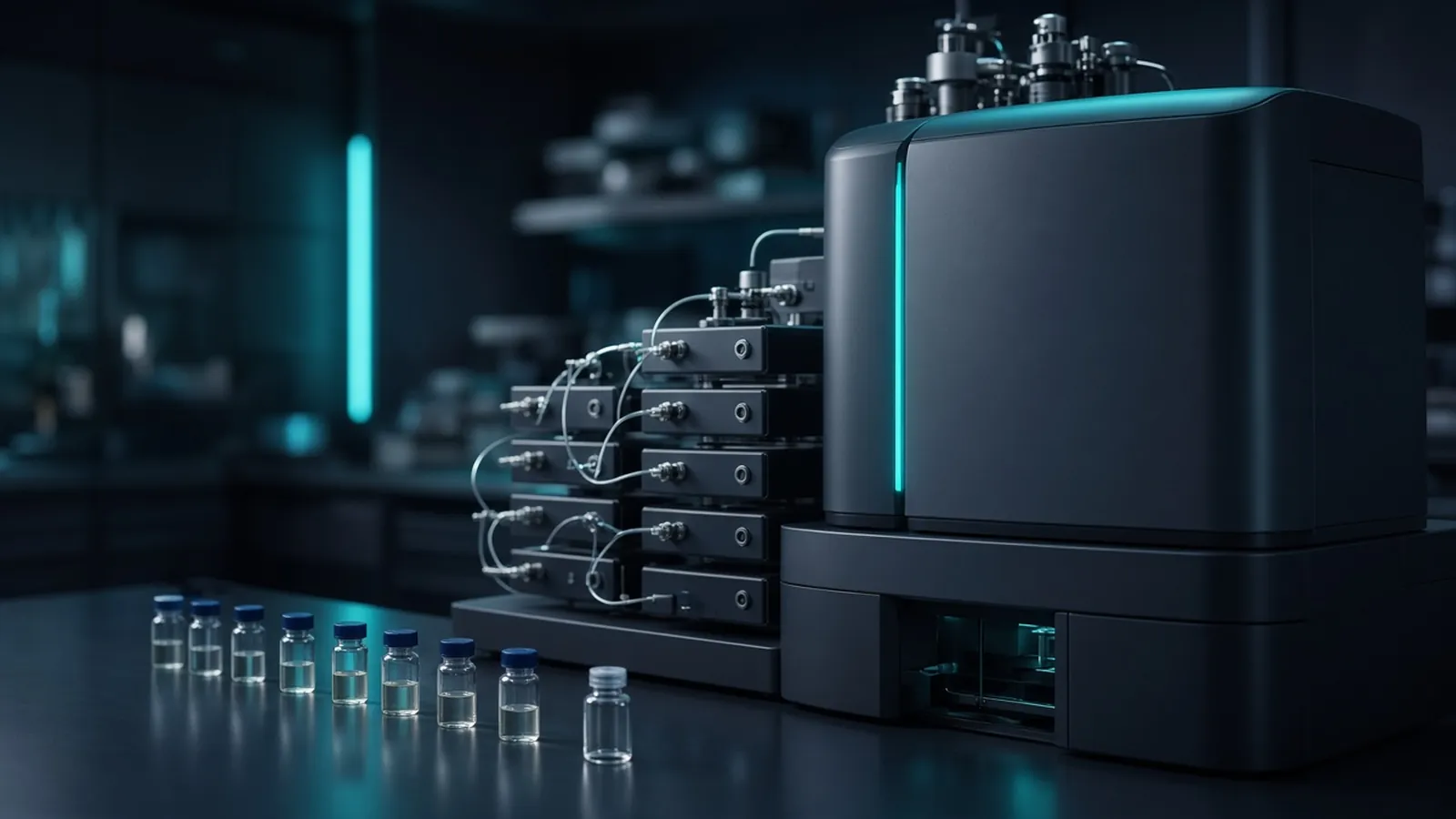
A peptide reversed-phase peak that fronts, splits, or arrives with the solvent disturbance is often an injection problem wearing a column's clothes. The decision this page supports is which check to run next, and which change will make the next injection interpretable. Where reversed phase sits among the other modes is covered in chromatography methods in life-science labs.
Hydrophobic retention in a water-rich start
Reversed phase holds peptides on a hydrophobic stationary phase, commonly a C18, C8, or C4 bonded silica, while the mobile phase starts rich in water and gains organic solvent. Acetonitrile is the organic solvent most peptide methods use, with a small acid modifier such as formic acid or trifluoroacetic acid. The acid keeps silanols and peptide side chains in a repeatable ionisation state. As acetonitrile rises, the mobile phase becomes a better solvent for the peptide, retention falls, and the peptide leaves. That is chromatography driven by a programmed change in solvent strength.
Retention time is the result of that programme on that column at that temperature and flow. A published minute mark without the gradient table is not a method. Pore size matters because a peptide from a large protein needs access to the bonded surface. Particle size and column length set pressure and narrowness. Follow the pressure rating. HPLC hardware delivers the flow. It does not choose the chemistry.
Ultraviolet detection near 214 or 220 nanometres sees the peptide bond and will show most peptides. Detection at 280 nanometres sees aromatic residues and will miss peptides that lack them. A mass spectrometer sees ions. Reporting those identifications in a proteomics study belongs to the standards community around HUPO. The precursor mass you expect can be checked against the sequence in UniProt. A retention time is still not that identification.
Symptom, likely cause, next check
Start with a blank gradient and a simple peptide standard in the starting mobile phase. If the standard is symmetrical and on time, the column and the pump are capable. If the standard is already split or fronting, stop. Fix the instrument before you interpret a digest.
When the standard is clean and the sample is ugly, compare the sample solvent with the starting mobile phase. A digest dried and redissolved in a high fraction of acetonitrile, or in dimethyl sulfoxide, is stronger than a gradient that starts at a few percent organic. The strong plug carries part of the peptide down the bed before the weak mobile phase can focus it. The peak fronts, breaks through near the void, or splits into a sharp early piece and a retained piece. The next check is dilution into the starting mobile phase, or evaporation and redissolution in a weaker solvent, then a smaller injection. ISO 8655-1 is a useful reminder that the pipette volume is a measured quantity with its own handling rules. Halving a bad solvent is still a bad solvent if it remains stronger than the start.
Tailing, with a slow return to baseline on the back of the peak, points somewhere else: overload, a tired column with extra silanol interactions, or a peptide that needs a different modifier. Reduce the load first. If the shape returns, the chemistry was fine and the mass was not. If a light load still tails, try the acid the column note prefers, or retire the column. Do not keep adding trifluoroacetic acid "until it looks better" on a mass-spectrometry method you still need to ionise.
No peaks at all, with a live baseline, is a short list. The lamp or the wavelength may be wrong for the peptides you have. The injection may not have happened. The gradient may never have become strong enough, so the peptides are still on the column. Extend the organic end on a blank-checked method, or strip the column as the insert allows, and watch the strip. A peak that appears only in a strong strip was retained. Your analytical gradient was too shy. A flat line through the strip means the peptides never arrived. Check the vial and the autosampler.
Everything eluting at the front, including a standard prepared correctly, means the column is not retaining. The starting organic fraction may be far too high, the phase may have been stripped by an out-of-range pH, or you are looking at a flow path that bypasses the bed. Confirm pH against the column window. A silica peptide column run for hours in strong base is a different object from the one you bought.
| Symptom | Likely cause to test first | Next check |
|---|---|---|
| Fronting, split peak, or breakthrough | Sample solvent stronger than the gradient start | Reinjection in starting mobile phase |
| Slow tail on a concentrated sample | Overload | Tenfold less mass |
| Slow tail on a light load | Extra interactions or a worn bed | Modifier and column history |
| No sample peaks, standard fine | Sample solvent, vial, or gradient too weak | Strip or extend, and watch |
| Standard and sample both early | Mobile phase composition or a dead column | Fresh start solvent and a second column |
Upstream cuts do not finish the map
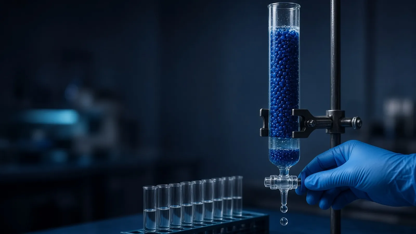
Affinity chromatography and ion exchange are common ways to simplify a protein before a digest, or to fractionate peptides when one reversed-phase dimension is not enough. They remove classes of molecules. They do not repeal co-elution on the reversed-phase column. If two peptides share hydrophobicity they can still draw one peak after a beautiful ion-exchange cut. Treat a single apex in a digest as unresolved response until mass spectrometry or a second gradient says otherwise.
Temperature belongs in the troubleshooting list because peptide retention moves with it. An oven that was off this morning and on yesterday will shift windows. Compare runs at the same set-point. A drifting baseline during the gradient can be absorbance of the acid or of the organic solvent itself. That is a mobile-phase property. Judge peaks from a blank subtracted only after you understand the blank, and keep the raw file.
Safety and research use
Acetonitrile, acids, and peptide samples are laboratory chemicals. Use the ventilation and waste stream your assessment names. Trifluoroacetic acid is corrosive. A research peptide map is not a clinical assay and not a purity release. Biosafety of the biological source is institutional. This page does not authorise a diagnostic claim from a chromatogram.
What to put in the enquiry
Name the peptide source, the detector, the acid you can accept, the sample solvent you currently use, analytical or preparative scale, and the failure you are seeing if you are replacing a column that misbehaves. Ask for bonded phase, pore size, particle size, dimensions, pH window, and pressure rating. The scientific instruments catalogue is the hardware class list. The specification goes on the quote request. If the peptides come from a purification campaign, the method can be discussed from the custom protein expression and purification reference without treating that page as a running service. ---
Questions from the bench
Why does a peptide peak split when the digest looked fine on the gel?
The gel reports size under denaturing conditions. The reversed-phase peak reports hydrophobic retention in a water and organic gradient. A split often means part of the injection rode through in a strong sample solvent while the rest focused at the column inlet. Dilute the sample into the starting mobile phase, or into a solvent weaker than that start, and inject again before you blame the digest.
Should formic acid or trifluoroacetic acid be in the peptide gradient?
Both are small acid modifiers used so the pH and the ionisation state stay defined. Trifluoroacetic acid often sharpens ultraviolet peaks and can suppress electrospray signal. Formic acid is the more common choice when a mass spectrometer is the detector. The pair is method-specific. Follow the column note and the detector you actually have, and do not swap the acid mid-campaign without re-checking retention and sensitivity.
A peptide map shows one clean peak for a region that should contain three peptides. What failed?
Co-elution is ordinary in a complex digest. One apex can hide neighbours the column did not resolve. Slow the acetonitrile gradient, change the column chemistry, or read the peak with mass spectrometry before you call the region pure. Affinity or ion-exchange work upstream can simplify the mixture. It does not guarantee that reversed phase will separate every remaining pair.
What should a peptide HPLC enquiry specify?
State peptide size range, analytical or preparative intent, detector (ultraviolet or mass spectrometry), preferred acid modifier, sample solvent, and the pressure the column must survive. Ask for chemistry, particle size, pore size, and dimensions. Send it with the quote request. A catalogue line that says HPLC does not name a peptide column.
References
Manufacturer names identify published method classes. Trademarks remain with their owners. Catalogue records on this site are independent references for enquiry. They are not a statement of inventory, distribution rights or a supply commitment. This page is educational. It is not medical advice, a diagnostic protocol or a biosafety approval.
Catalogue
Related products and categories
These links follow the subject of the article into published manufacturer references. A listing is a reference for an enquiry, not a statement of stock or distribution rights.
Continue in this cluster
Related reading
 Chromatography methods in life-science labsHow life-science chromatography separates molecules on a stationary phase, and what an HPLC or column peak does not prove about purity.A glossary of chromatography termsUse chromatography terms with their real limits: retention, resolution, void, and a peak that is only a detector response.
 Affinity and ion-exchange protein purificationHow affinity capture and ion exchange pull a protein from a lysate, and why each collected fraction still needs an identity check.