
protocol overview
SDS-PAGE for protein size estimates
Plan an SDS-PAGE size estimate: stacking and resolving gels, SDS and reduction, apparent mass, and a ladder you do not read past.
- Author
- EVRINTH Editorial Team
- Published
- 8 October 2026
- Updated
- 8 October 2026
- Reading time
- 9 min
SDS-PAGE is how a laboratory turns a protein mixture into a lane of bands sorted mostly by polypeptide length. The estimate you write down is an apparent mass: where the band sat relative to a ladder on that gel, after detergent, charge and usually a reducing agent and heat had done their work. This page is the staged overview of that estimate. How to read the finished lane, including smears and material trapped in the well, is reading a protein gel. DNA sorting on agarose is a different method, agarose gel electrophoresis for DNA. Nothing here is a clinical protein electrophoresis or a licence to cast acrylamide.
Gels, ladders and general molecular supplies are enquiry items in the molecular biology catalogue. Put the mass window and whether you cast or buy precast on the quote request.
Who needs an apparent mass
Expression checks, purification fractions and a lysate you are about to blot all need a size window before they need a name. The decision is whether a species near the designed mass is present, whether neighbours were removed, and whether the sample entered the gel. The sequence mass is the plan. UniProt is a public place to confirm an unmodified mass and known processing. The plasmid map wins if you added a tag the database entry lacks.
Say "apparent mass" in the notebook. A band that migrates with the 50 kilodalton rung was not weighed. It travelled like that rung under these conditions.
What the detergent, the reducer and the mesh each do
Sodium dodecyl sulfate binds along most polypeptides and swamps their own charge with a negative coat roughly in proportion to length. In the field they move toward the anode, and the polyacrylamide mesh sieves them so longer chains lag. That is why a denaturing gel can be read as a size estimate at all.
A reducing agent of the dithiothreitol or beta-mercaptoethanol class breaks disulfide links so subunits and knotted domains can unfold. Heat finishes that unfolding for many soluble proteins. Leave out the reducer and a disulfide-linked species can run at a different apparent mass. Leave out the heat and some domains stay folded enough to bind SDS unevenly. Membrane proteins and very acidic or basic proteins are the classic exceptions even when you do both. Incomplete reduction is a result. It is described as a clue in the reading guide, not as a broken power supply.
The Laemmli-style gel is two layers. A short stacking gel, typically a low acrylamide percentage at a lower pH, sits on a resolving gel of the percentage you chose. The running buffer is a Tris-glycine system with SDS. At the stacking pH the ions arrange so proteins compress into a thin zone before they enter the resolving mesh. That compression is why a sloppy load can still become a sharp band. It is not magic, and it fails if the layers were poured so the boundary is ragged, or if the sample never reached the stack because it aggregated in the well.
Percentage is a concept tied to pore size. A higher acrylamide percentage tightens the mesh, resolves smaller polypeptides, and can exclude large ones so they sit at the top of the resolving gel or never leave the stack. A lower percentage lets large chains enter and separate, and it lets small ones run into the dye front. Gradient gels span a wider window in one slab. Many laboratories plan a higher percentage for a small chain, something near the middle of the usual cast range for an ordinary soluble protein, and a lower percentage for a large one. Follow the precast cassette you actually bought, or the institutional casting SOP if you pour. This page deliberately has no gram table, no initiator volumes and no bis-acrylamide ratio.
Loading dye in the sample buffer adds density and a tracking colour. The dye front is where the smallest species and the free dye collect. It is not a mass rung. Stop the gel while the front is still on the plate if small proteins matter.
A workflow with the branches
Look up the mass, including tags and cleavage scars. Decide reduced or non-reduced on purpose, and write it on the gel. A comparison between those two lanes is a disulfide question. A single reduced lane is a size question. Mix the sample with SDS sample buffer of the class your laboratory uses. Apply heat if that buffer and that protein call for it. Cool the tube before you load. A film of condensation is not an extra sample.
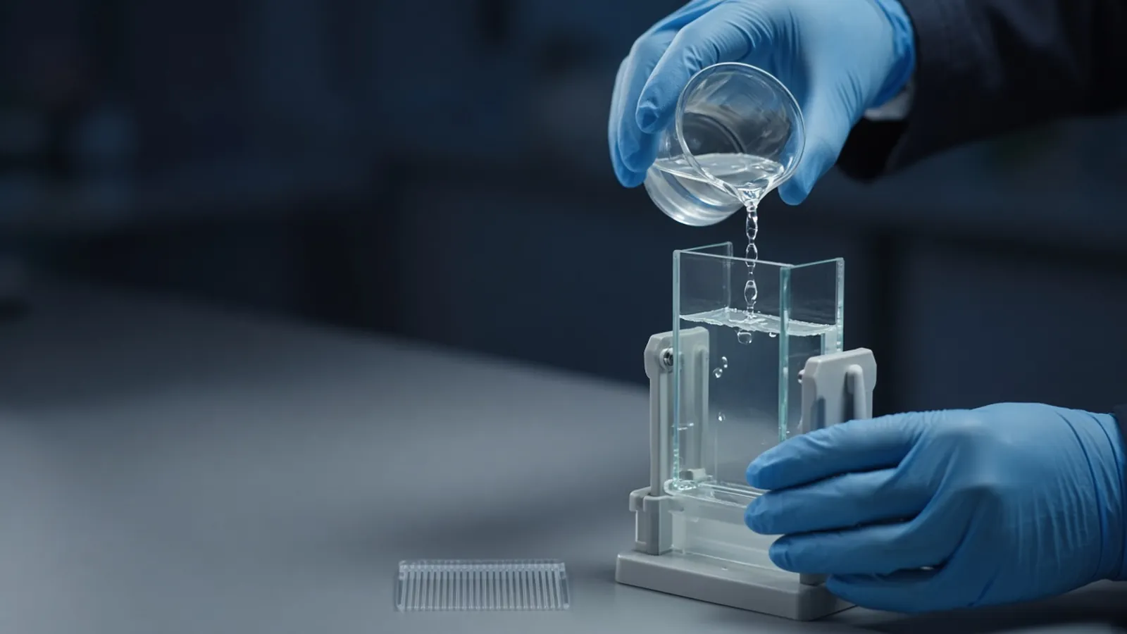
Cast only if your SOP allows it. The photograph is that step and nothing wider: gloved hands pouring solution between glass plates held in a casting stand. There is no face in that frame, and there is no second observer to invent. Keep the plates clean, the spacers matched, and the resolving gel set before the stack goes on. A leak is a chemical spill of monomer until it has polymerised. Precast cassettes skip that hazard and still need the percentage on the cassette, not the percentage you remember from a hand-cast gel last year.
Load a ladder that brackets the expected apparent mass. Prestained markers let you watch the run and, later, a transfer. Their dyes shift apparent size, so they are the wrong ruler when the estimate itself is the result. Unstained markers, stained afterwards with the same total-protein stain as the samples, are the fairer comparison. Do not draw the line past the last rung you can see.
Run until the window you care about has opened, at a field that does not cook the plates. Smile-shaped lanes mean the centre was hotter than the edges. A western blot can follow, and the transfer is its own experiment, described from gel to membrane in the cluster's blot guide. An agarose habit does not transfer. Protein percentage, protein ladders and protein stains are the record.
After staining, describe the lane before you name the protein. A sharp band near the plan supports an abundant species of about that apparent mass. Extra bands are other polypeptides until an antibody, an activity, or mass spectrometry says otherwise. The Human Proteome Organization's public materials are a reminder that identity claims sit in a wider quality conversation. They are not a gel recipe.
| Choice | What it is for | What it does not give you |
|---|---|---|
| SDS plus reducing agent and heat | A denatured chain, apparent mass | Native shape, activity, or a certified mass |
| Non-reducing lane beside it | Whether a disulfide changed the species | A full native-complex map |
| Higher percent acrylamide | Smaller proteins spaced apart | A fair view of a very large chain |
| Lower percent acrylamide | Large proteins able to enter | Small proteins kept off the dye front |
| Unstained ladder | Size comparison after the same stain | A number outside its rungs |
| Prestained ladder | Watching the run and the transfer | A precise apparent mass |
When the estimate fails
No bands, including the ladder, means the field, the stain, or the load failed. Check those before you rewrite the construct. Protein trapped in the well can be aggregated, insufficiently denatured, or too large for that percentage. A smear from well to dye can be degradation or gross overload. Repeat with less protein before you invent an isoform. A band far from the sequence mass can be a real modification. It can also be the wrong polypeptide. The reading guide and a later blot are how you refuse the quicker story.
An oxidised reducing agent in an old bottle leaves disulfides intact, so the "reduced" lane is not reduced. If a harsh smell is your only quality check, you do not have a check. Follow the laboratory's rule for how long that bottle stays in service.
Safety and research limits
Acrylamide monomer is a neurotoxin before it polymerises. Casting, including the pour in the photograph, belongs under the institutional SOP: who may do it, what gloves and waste path are required, and how leftover monomer is discarded. Polymerised gels are still chemical waste. Reducing agents irritate. SDS is a chemical hazard of its own class. This article does not include a recipe, and it does not authorise the work.
Biological samples follow the biosafety level of their source. Detergent does not reclassify a culture. The WHO Laboratory biosafety manual, 4th edition is background, not a permit. A research gel supports a biochemical note about that sample. It is not a diagnostic electrophoresis.
Warm rooms and gels that set too fast
Hand-cast gels in a hot room polymerise faster. The pour between the plates has to happen before the solution sets in the vessel, which is an SOP problem of timing, not a reason to skip protection. Do not raise the running voltage to finish before the next power cut. Hot plates smile, and a smiled ladder cannot support an apparent mass. If the building supply is unreliable, prefer a run you can watch to completion. A gel that sat mid-run without a field lets bands diffuse. Repeat it. Precast gels stored warmer than their label allows are a separate failure. Follow the storage on the box.
What to put in an enquiry
State the apparent-mass window, whether you need precast cassettes or your institution casts, the ladder style (prestained for tracking, unstained for the estimate), and whether the gel must later transfer for a blot. Name reducing-agent class only as a sourcing category your SOP already allows. The nucleic acid analysis pathway is the neighbouring route when the same bench also orders reagents for the DNA work beside this gel. Ask whether a quotation is possible. Ask for the percentage and the ladder range in writing. Do not accept a cassette whose percentage is missing from the description.
Set up an SDS-PAGE estimate you can defend
- 01Write the sequence mass and the chemistryRecord the unmodified mass from the construct you expressed, including tags, and whether the prep will be reduced and heated. Apparent mass is what the gel will show.
- 02Choose a resolving percentage for that windowUse a higher acrylamide percentage when the chain is small and a lower percentage when it is large. Treat the number as a concept matched to the gel you cast or buy, not as a recipe copied from another laboratory.
- 03Bracket the band with a ladder on that gelLoad a marker that flanks the expected apparent mass, using prestained ladders to watch the run and unstained markers for the fairer size comparison. Do not report a mass outside the rungs.
- 04Separate size from identityA band near the plan supports an abundant species of about that apparent mass. Name the protein only after an orthogonal check such as a blot, activity, or mass spectrometry, and read the lane with that limit in mind.
Questions from the bench
Why does my protein not match the mass calculated from the sequence?
SDS-PAGE reports apparent mass under the conditions you used. Glycosylation, other modifications, an unusual composition, a tag, incomplete unfolding, or a prestained ladder that itself migrates oddly can all shift the band. Compare with an unstained ladder inside its range, and look up known processing before you edit the sequence to match the gel.
Do I have to boil every sample?
Heat is part of a typical denaturing prep, and many sample buffers ask for a short incubation near boiling or in a hot block. Some membrane proteins aggregate when boiled and then sit in the well. Follow the sample buffer and the protein class. Skipping heat without a reason leaves folded species that will not match a fully denatured ladder.
Can I pour this from a recipe I found online?
Unpolymerised acrylamide is a neurotoxin. Who may weigh it, pour it and dispose of it is an institutional SOP, not a blog recipe. The photograph shows gloved hands pouring solution between glass plates in a casting stand, which is that controlled step. Precast gels are the alternative class when your laboratory does not cast.
Is a band at the right apparent mass enough to start a western blot claim?
It is the separation half of the claim. A western blot still has to transfer that pattern and mark it with an antibody whose controls survive. An agarose gel of a PCR product is a different matrix and a different molecule. Do not move a DNA-ladder habit across to a protein mass.
References
Manufacturer names identify published method classes. Trademarks remain with their owners. Catalogue records on this site are independent references for enquiry. They are not a statement of inventory, distribution rights or a supply commitment. This page is educational. It is not medical advice, a diagnostic protocol or a biosafety approval.
Catalogue
Related products and categories
These links follow the subject of the article into published manufacturer references. A listing is a reference for an enquiry, not a statement of stock or distribution rights.
Continue in this cluster
Related reading
 Agarose gel electrophoresis for DNAHow agarose percentage, buffer and a size ladder turn a DNA sample into a band, and what that band does and does not prove about identity.A glossary of electrophoresis termsDefine anode, cathode, sieving, mobility, stacking, resolving, electroendosmosis, Rf, and leading dye for an honest gel reading.
 Blocking buffers and background signalChoose milk or BSA and a wash that fits the antibody, then decide whether speckles are sample signal or block, secondary, or a dried membrane.