
application
Troubleshooting a blank western blot
Reason through a blank western in order: load, transfer, membrane face, antibody, azide, substrate, washes, then a true absence.
- Author
- EVRINTH Editorial Team
- Published
- 8 October 2026
- Updated
- 8 October 2026
- Reading time
- 8 min
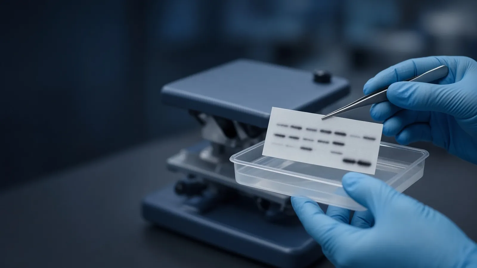
A blank western blot is a white or evenly grey membrane where a band was expected. The useful decision is which step failed, in an order that does not skip to "the antigen is absent". This page is that order. The photograph shows gloved hands lifting a membrane with forceps over a buffer tray, with a rocking shaker behind. That handling, keeping the sheet wet and the gel-facing side known, is where several blank blots are made or avoided. The full path from gel to signal sits in western blot from gel to membrane. A DNA gel is a different kind of evidence, read in agarose gel electrophoresis for DNA.
Stop troubleshooting and repeat the biology only when a positive control on the same sheet develops and the test lane is still blank for a reason you have not already disproved. Until then, change one variable.
No protein was loaded
The first blank is an empty well. The lysate precipitated and was left in the tip. The tube was the wrong fraction. The volume was buffer. A loading dye that ran down the lane only proves the electrical field moved small ions. It does not prove polypeptide entered the resolving gel.
Stain the gel after transfer, or stain a sister lane that you did not blot. Protein still in the gel was loaded and did not leave. An empty lane beside a normal molecular weight ladder means the sample never arrived. Load a known mass before you incubate another antibody.
Transfer failed
Proteins become a blot only if they leave the gel and stay on the transfer membrane. No current, a short time, a bubble, a dry cassette, or a sandwich in the wrong order all produce a blank. A prestained ladder is the witness. If those coloured bands are still in the gel, the field did not move them. If they are on the paper stack and the membrane is white, they moved the wrong way. If they are on the membrane, transfer of at least that size range happened.
Large proteins are the ones left behind when the clock is kind to a small target. Small proteins are the ones that pass through a large pore. A second membrane behind the first, or a total-protein stain such as Ponceau, separates "never left" from "went through". A blank Ponceau ends the antibody conversation for that sheet. Activate PVDF in the solvent the membrane maker names before it will wet.
The membrane was flipped
Protein binds to the face that met the gel. Probe the other face and the signal is weak or gone, even though a faint prestain may show through. Forceps, as in the photograph, lift the sheet without scraping that face. A cut corner, written down, is the memory. A membrane flipped during a wash looks like an antibody failure on the next development.
Stack order is the earlier version of the same mistake. Filter paper, gel, membrane, filter paper, with the field pointed so protein moves into the membrane. Reverse the cassette and the membrane was never in the path.
The antibody pair does not match
A mouse primary needs a secondary raised against mouse. A rabbit primary with an anti-mouse secondary is a blank with extra incubations. Host species is on the vial. So is the clone or the catalogue identity you must write in the notebook. A primary against a different isoform, or against a tag your construct does not carry, binds nothing in this lysate and can still be a good antibody. Check the expected mass and known processing on UniProt, then check the construct you actually expressed.
Include a strip that sees only the secondary. A band there is background. If that strip and a positive lysate are both blank, the primary, the transfer, and the detection are still open. Change one of them.
Azide killed the HRP secondary
Horseradish peroxidase is the enzyme on many chemiluminescent secondaries. Sodium azide inhibits it. Antibodies are often stored in azide so microbes do not grow. That preservative, carried into the antibody bath or into a wash, can extinguish the enzyme after the primary has done its job. The membrane is blank. The proteins are there. A fluorescent secondary does not use that enzyme and will not fail for this reason, which is a useful branch if you can detect another way.
If the primary vial says it contains azide, keep that dilution away from the HRP step or follow the sheet's instruction for an HRP-compatible dilution. Do not "fix" a blank by adding more substrate on top of an inhibited enzyme. Confirm the detection class first.
The substrate was exhausted
Chemiluminescent substrates are mixtures with a life after they are combined, and a life on the shelf before that. An old bottle, a bottle left in the light, or a working reagent mixed and then forgotten will not glow even on a blot that has worked for months. The test is a known positive, or a dot of the secondary on a spare corner of membrane, developed with the same mix. If that dot stays dark, the substrate is finished. Mix a fresh preparation when the sheet is in the tray, and follow the timing on the substrate sheet. Alkaline-phosphatase substrates will not rescue an HRP secondary. Match the enzyme.
The washes stripped the signal
Washes remove antibody that is only resting on the membrane. A wash harsher than the antibody tolerates, or one that runs while you are elsewhere, can lift a weak specific signal too. The positive control fades with the test lane when the wash is the cause. High background points the other way. Change the wash last, back toward a condition that still shows the positive control.
A membrane that dried at the edge during a long wash stains in patches and can hide a real band in a dark field. The rocking shaker in the photograph only helps if buffer still covers the whole sheet. Dried PVDF is a storage choice some protocols allow at the end. It is a different event from drying in the middle of the primary incubation.
Only then ask whether the antigen is absent
When the ladder transferred, Ponceau shows the test lane, the secondary matches, azide is out of the HRP path, the substrate develops a positive lysate, and the washes spare that band, a blank test lane means this antibody did not mark the antigen in this fraction. The protein may sit in a compartment the lysis never opened, or the epitope may be the splice form you did not load. UniProt lists known processing. The Human Proteome Organization's public materials remind you that an identity claim wants more than one method when the conclusion matters.
| Step in order | What a pass looks like | If it fails, stop here |
|---|---|---|
| Protein loaded | Sister lane or post-transfer gel stain shows sample | Reload a known mass |
| Transfer | Prestained ladder on the membrane, Ponceau in the lane | Fix field, time, bubbles, or pore |
| Face and stack | Gel-facing side known, bands not on the cushion paper | Re-transfer with the stack marked |
| Antibody pair | Host species matches, positive lysate can work | Correct the secondary or the primary identity |
| Azide and HRP | Detection class tolerates the preservative in the vial | Remove azide from the HRP path |
| Substrate | A dot of secondary still develops | Fresh substrate, matched enzyme |
| Washes | Positive band survives the wash you used | Return to a milder wash |
| Antigen | All rows above passed, test lane blank | Report absence in this fraction only |
Safety, heat, and a cut in the mains
Methanol for PVDF, and unpolymerised acrylamide in the gel you came from, are chemical hazards under your institutional rule. Lysates follow the biosafety level of the cells. This page does not authorise a clinical blot.
In a hot laboratory an ambient antibody incubation runs warmer than the protocol assumed, and background rises. A power cut during transfer leaves a partial ladder. Keep buffer over the membrane. That is the point of the tray in the photograph.
What to send when the blank is why you are enquiring
EVRINTH can take a sourcing question. Name the target and the expected mass, the host of the primary, the membrane and pore class, whether detection is HRP or fluorescent, and which of the eight steps already passed. The molecular biology catalogue is the enquiry list, and the quote request is where the requirement goes. The nucleic acid analysis pathway is the neighbouring workflow when the same project also measures a transcript. A blank blot and a missing PCR band are not one fault. Ask whether a quotation is possible. Send the step that failed, not only the protein nickname.
Questions from the bench
Should I call a blank lane a biological negative?
Only after the controls that can still light up have done so. A known positive lysate on the same membrane, a molecular weight ladder that transferred, a total-protein stain in the test lane, a matched secondary, a live substrate, and washes that spare the positive band are the gate. Until that gate is passed, a blank is a method result.
What does sodium azide do to an HRP secondary?
Sodium azide, often present so an antibody solution keeps, inhibits horseradish peroxidase. A working dilution that carries azide into the detection can kill the enzyme and leave a white membrane even when the primary bound. Fluorescent secondaries are a different detection class and do not fail by that particular poison. Follow the antibody note before you mix a stored primary into an HRP blot.
How do I tell a flipped membrane from a failed transfer?
A prestained ladder that is visible on the membrane shows that protein moved and that you are looking at a face that received it. A ladder still in the gel, with a blank membrane, means the field, the time, or the stack never delivered protein. A ladder on the filter paper you thought was only a cushion means the stack was reversed. Mark the gel-facing side with a cut corner before the stack comes apart.
The loading dye ran, but the blot is blank. What does that clear?
The loading dye shows that the gel electrophoresis itself moved ions. It does not show that protein left the gel, that the membrane was the right way up, or that the antibody pair can work. Keep the dye observation, then go to a total-protein stain of the membrane before you order a new primary.
References
Manufacturer names identify published method classes. Trademarks remain with their owners. Catalogue records on this site are independent references for enquiry. They are not a statement of inventory, distribution rights or a supply commitment. This page is educational. It is not medical advice, a diagnostic protocol or a biosafety approval.
Catalogue
Related products and categories
These links follow the subject of the article into published manufacturer references. A listing is a reference for an enquiry, not a statement of stock or distribution rights.
Continue in this cluster
Related reading
 Agarose gel electrophoresis for DNAHow agarose percentage, buffer and a size ladder turn a DNA sample into a band, and what that band does and does not prove about identity.A glossary of electrophoresis termsDefine anode, cathode, sieving, mobility, stacking, resolving, electroendosmosis, Rf, and leading dye for an honest gel reading.
 Blocking buffers and background signalChoose milk or BSA and a wash that fits the antibody, then decide whether speckles are sample signal or block, secondary, or a dried membrane.