
guide
Western blot from gel to membrane
How a protein moves from an SDS gel onto a membrane, how antibodies create a band, and which controls separate transfer failure from a real signal.
- Author
- EVRINTH Editorial Team
- Published
- 8 October 2026
- Updated
- 8 October 2026
- Reading time
- 8 min
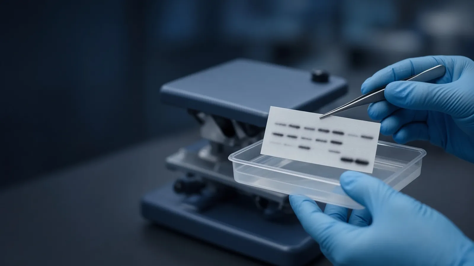
A western blot asks whether a protein of roughly a certain size is present in a lysate, and whether an antibody will mark it after the proteins have been stuck to a membrane. The gel does the sorting. The transfer makes a stable copy of that pattern. The antibody does the identification, within the limits of that antibody. This page follows that path and the controls that stop a blank membrane from being called a biological negative. It is a research explainer, not a clinical immunoassay and not a vendor protocol.
DNA gels answer a different question with a different matrix. Read them in agarose gel electrophoresis for DNA. Blotting reagents and general molecular supplies are enquiry items in the molecular biology catalogue. Attach the target and the detection class to the quote request.
From a crowded lysate to a lane
Cells contain thousands of proteins. Sodium dodecyl sulfate denatures most of them and coats them with negative charge roughly in proportion to length, so they migrate through a polyacrylamide gel more by mass than by their native shape. That approximation fails for heavy glycosylation, for some very basic proteins, and for proteins that never fully unfolded. A band "at 50 kilodaltons" means "migrated with the 50 kilodalton marker under these conditions", not "the polypeptide is certainly 50 kilodaltons and unmodified".
A molecular weight ladder belongs in an outer lane. Prestained ladders let you watch the transfer. The dye changes the apparent size, so use them to track orientation. Use an unstained mass standard when a precise size claim is the point of the figure.
Unpolymerised acrylamide is a serious chemical hazard. Cast gels inside the practice your institution already set, and treat leftover monomer as chemical waste. This article does not include a recipe. Power supplies and cracked plates are the everyday physical risks.
Transfer is its own experiment
Proteins do not become a blot until they leave the gel and stay on a membrane. Wet-tank transfer and semi-dry transfer are the common classes. Both use an electric field and a stack that must be in the right order, with no air bubbles to cast a blank oval. The field has a direction. Reverse it and the proteins travel into the filter paper you thought was only a cushion.
Nitrocellulose wets in aqueous buffer and is often enough for abundant proteins. PVDF is tougher and binds more protein per area, and it typically needs a brief activation in methanol, or the solvent the membrane maker specifies, before it will wet in aqueous buffer. A PVDF sheet that was never activated looks white and stays empty. That is a transfer failure, not a knockout.
Size decides the timing. Large proteins move slowly and are the ones left behind if you shorten the transfer to be kind to a small protein. Small proteins can pass through the membrane entirely, especially if the pore size is large and the run is long. A second membrane behind the first, or a total-protein stain, catches that. Ponceau and similar reversible stains show that protein arrived. They do not identify it. A beautiful Ponceau with no antibody signal points at blocking, the antibody, or the detection. A blank Ponceau points at the transfer or the load.
Antibodies and the background they live in
Blocking covers the membrane so the antibody does not stick everywhere. Milk, serum albumin and commercial blockers are classes, not universal solvents. A phospho-specific antibody can pick up phosphorylated milk proteins and look filthy. Match the blocker to the antibody note, and keep that choice when you compare blots.
The primary antibody should name its host species, its clone or catalogue identity, and the dilution you actually used. Store it as the supplier's label says. A working dilution left warm all afternoon is a different reagent. The secondary antibody has to recognise that host. A mouse primary with an anti-rabbit secondary is a blank blot with extra steps. Include a strip that skips the primary. Any band there belongs to the secondary, the detection reagent, or the block, and it cannot be claimed as the target.
Washes remove antibody that is merely resting on the membrane. Too little washing raises background. Too much, or a detergent that is harsher than the antibody tolerates, can strip a weak specific signal. Change one variable at a time when you are debugging. Detection with an enzyme and a chemiluminescent substrate, or with a fluorescent secondary, both saturate. Expose a series, or set the imager inside a range you have checked, before you rank lanes. A loading control such as a cytoskeletal protein is traditional and sometimes dishonest, because treatments change those proteins. A total-protein stain of the same membrane is often the fairer loading record. The Human Proteome Organization's public materials are a reminder that protein identity claims sit inside a wider quality conversation. They are not a blot protocol.
| Control | What it is for | Failure you should believe |
|---|---|---|
| Prestained ladder after transfer | Orientation and the fact of transfer | Ladder absent on the membrane means proteins likely stayed in the gel or the stack was reversed |
| Total-protein stain | Load and transfer across the lane | Blank membrane, antibody not worth blaming yet |
| Secondary only | Reagent background | Bands that appear without primary are not the target |
| Lysate known to contain the protein | The reagents can succeed | A failed positive lysate indicts antibody, detection or transfer |
| Lysate known to lack the target | Specificity | A band in the negative lysate is cross-reactivity or contamination |
Failure modes
High background with a visible specific band can be too much antibody, a dirty dish, a blocker that does not suit the antibody, or a membrane that dried between steps. Uneven background often means uneven shaking or bubbles. A rocking platform only helps if the buffer actually moves over the whole sheet.
No specific band, with good total protein and a working secondary pair, points at the primary antibody, the epitope, or a target that is not in this fraction. Membrane proteins and nuclear proteins are easy to lose in a sloppy lysis. Check that the lysate method matches the compartment you care about. A band at an unexpected size can be a real isoform, a cleavage product, or a cross-reactive protein. Look up known processing on UniProt and then test, rather than cropping the lane to the size you hoped for.
Room conditions and safety
Methanol used to activate PVDF is flammable and toxic. Acrylamide monomer is neurotoxic before it polymerises. Follow local chemical rules. Biological lysates follow the biosafety level of the cells, not the fact that you added detergent.
In a hot laboratory, antibody incubations drift warmer than the "room temperature" written in a protocol from a milder climate. Background rises, and some antibodies do not appreciate it. A cooled room or a shortened incubation is a documented change, not a secret. A power cut during transfer leaves a partial pattern. Do not treat that membrane as quantitative. Finish or repeat the transfer. Do not invent the missing minutes.
Humidity and air drafts dry the edge of a membrane while the centre is still wet. Dried patches stain darkly and ruin the comparison. Keep the sheet wetted during washes. Some PVDF protocols allow a fully processed membrane to be dried for storage. That is a deliberate end step, not the same as drying halfway through the primary incubation.
What an enquiry should carry
EVRINTH can take a sourcing question. Name the target, the expected mass, the host species of the primary antibody if you already know it, the membrane class, and whether detection is chemiluminescent or fluorescent. The nucleic acid analysis pathway is a neighbouring workflow when the same project also measures the transcript. A blot and a nucleic-acid result are not interchangeable evidence. Ask whether a quotation is possible. Do not send only a protein nickname and expect a single antibody to be the obvious choice. Isoforms and modification states are part of the specification.
Take a protein from a gel to an interpretable blot
- 01Separate, then prove the protein left the gelRun an SDS gel with a molecular weight ladder in a lane you can track after transfer. Stain the membrane for total protein, or check a reversible stain, before you celebrate an antibody band.
- 02Match the membrane to the proteinActivate PVDF if that is the membrane class you are using. Choose transfer time with the size of the target in mind so large proteins actually move and small proteins do not pass through.
- 03Block, probe and keep a secondary-only laneBlock spare membrane, incubate the primary antibody in its stated conditions, and include a lane or a blot piece that sees only the secondary reagent. Wash enough that fresh buffer still comes off clean.
- 04Call the band only if the controls agreeA mark at the expected size is a candidate. A missing loading control, a blown-through transfer, or a secondary-only band at the same place withdraws the claim. Confirm identity with an orthogonal check when the conclusion matters.
Questions from the bench
Why is there no band when the gel looked fine?
The protein may never have left the gel, or it may have passed through the membrane. A total-protein stain of the membrane distinguishes those two failures from a bad antibody. Antibody problems remain possible when the stain shows protein and the specific signal is still absent. Check the host species of the secondary against the primary you actually used.
Does a band at the predicted mass prove the antibody is specific?
It is consistent with specificity and compatible with a cross-reactive protein of similar size. A cleaner argument uses a lysate that lacks the target, a competing peptide if the antibody was raised that way, or a second method such as mass spectrometry. UniProt is a public place to confirm the expected mass and known processing, not a substitute for that experiment.
Can I compare band darkness between lanes as a quantity?
Only inside the linear range of the detection, with loading controlled by total protein or by a loading control you have shown does not change with the treatment. Saturated films and over-exposed cameras compress real differences. Most casual blots are better at presence and approximate size than at a fold change.
Is a western blot the same kind of evidence as an agarose gel?
No. An agarose gel sorts nucleic acids by how they thread a matrix. A western blot sorts proteins by approximate mass and then asks an antibody which band to darken. The ladder, the failure modes and the claim are all different. Use the agarose note for DNA and this note for protein.
References
Manufacturer names identify published method classes. Trademarks remain with their owners. Catalogue records on this site are independent references for enquiry. They are not a statement of inventory, distribution rights or a supply commitment. This page is educational. It is not medical advice, a diagnostic protocol or a biosafety approval.
Catalogue
Related products and categories
These links follow the subject of the article into published manufacturer references. A listing is a reference for an enquiry, not a statement of stock or distribution rights.
Continue in this cluster
Related reading
 Agarose gel electrophoresis for DNAHow agarose percentage, buffer and a size ladder turn a DNA sample into a band, and what that band does and does not prove about identity.A glossary of electrophoresis termsDefine anode, cathode, sieving, mobility, stacking, resolving, electroendosmosis, Rf, and leading dye for an honest gel reading.
 Blocking buffers and background signalChoose milk or BSA and a wash that fits the antibody, then decide whether speckles are sample signal or block, secondary, or a dried membrane.