
comparison
Detectors UV fluorescence and mass spec
Choose UV, fluorescence, or mass spectrometry by what each detector can report, and what it still cannot identify alone.
- Author
- EVRINTH Editorial Team
- Published
- 8 October 2026
- Updated
- 8 October 2026
- Reading time
- 7 min
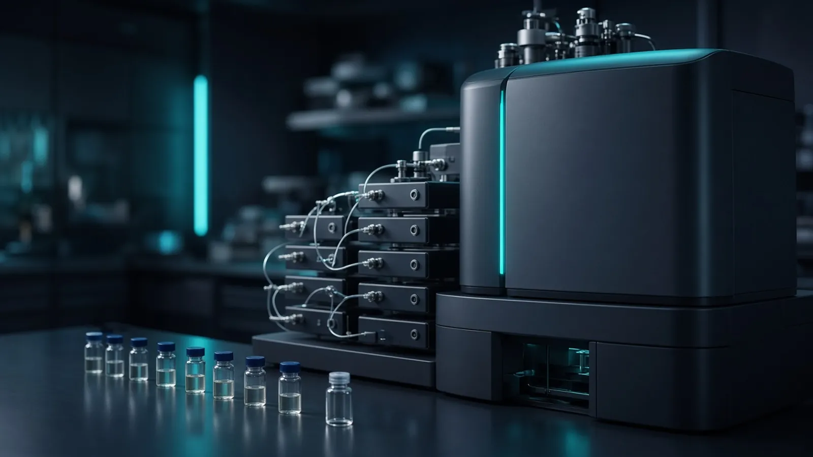
Ultraviolet absorbance, fluorescence, and mass spectrometry answer different questions about the same column effluent. The decision is which question you have, and which claim you must not let the trace make. The column work upstream of the flow cell is set out in chromatography methods in life-science labs.
Three responses, one elution
Chromatography separates. The detector transduces a property of whatever co-elutes in the cell at that moment. Ultraviolet absorbance measures how much light at one wavelength fails to get through the cell. Fluorescence measures light given back at a longer wavelength after excitation. A mass spectrometer measures ions. If two compounds leave the column together, all three detectors see a mixture, and only the mass spectrometer has a chance of showing that the mixture has more than one mass. Even then, suppression can hide the quieter ion.
Path length and concentration connect absorbance to response. A longer cell sees more of a dilute peak and saturates sooner on a preparative stream. Fraction collection after the cell must account for the cell volume and the tube to the drip. Column volume still sets the separation. The detector does not improve a separation that happened, or failed, upstream.
Ultraviolet absorbance
Use ultraviolet detection when the analyte has a chromophore and you want a robust, quantitative trace. Proteins and peptides are the everyday case. Aromatic residues dominate near 280 nanometres. Peptide bonds dominate near 214 to 220. A protein with few aromatic residues can be abundant and nearly invisible at 280. UniProt shows the sequence you can inspect for tryptophan and tyrosine before you declare a purification empty. Buffer choice is part of the detector. Imidazole, trifluoroacetic acid, and some organic solvents absorb in the low ultraviolet and lift the baseline as a gradient proceeds. A peak on a sloping baseline needs a blank before you integrate it.
Absorbance does not identify. A standard at the same retention is consistency, not proof. An impurity with a stronger chromophore can dominate the area while contributing little mass. Report area as quantity only inside a calibration made with the real compound. Extrapolating a curve is how detectors invent precision.
Fluorescence
Fluorescence is the selective optical detector. Native fluorescent residues, cofactors, or a deliberate label can make the analyte bright against a dark matrix. Set excitation and emission from the fluorophore you actually have, not from a neighbouring filter that "usually works". Quenching by the mobile phase, the dissolved oxygen, or a high concentration can flatten the response just where the ultraviolet channel still climbs. The two detectors in series are a useful argument: a peak in both is stronger than a peak in one, and a peak in only one tells you which property you lack.
A label changes the molecule. The retention you measure is the labelled molecule's retention. Quantifying it as if it were the unlabelled analyte requires a recovery you measured. Fluorescence of a collected fraction in a plate reader is a different optical path from the flow cell. Do not expect the numbers to match without a shared calibration. Neither reading names an unknown contaminant that happens to carry a similar dye.
Mass spectrometry
Mass spectrometry is the detector that can support an identification, and the one most often over-trusted. A mass-to-charge ratio consistent with the sequence, plus fragment ions that match, is strong research evidence. Reporting it well is the kind of practice HUPO exists to improve. A single mass that several peptides could explain is a weaker claim. Co-elution still mixes spectra. Ion-exchange or another chromatographic dimension is how you present the spectrometer with fewer neighbours, not a luxury after the fact.
The spectrometer does not see what does not ionise. Neutral, salty, or suppressed analytes can be major by absorbance and absent from the ion chromatogram. Salt from an ion-exchange fraction and imidazole from an affinity elution suppress and contaminate sources. Exchange the buffer before the vial. Solvents and acids have to be chosen for the source: formic acid is a common electrospray choice, and trifluoroacetic acid often sharpens ultraviolet peaks while suppressing spray. The detector choice feeds back into the mobile phase. Decide it before you lock the gradient.
A total-ion chromatogram is not an ultraviolet trace and will not show the same relative heights. Extract an ion if you are following one mass. Keep the optical channel if you need to see the things that did not ionise. The fraction you collect is still a mixture of whatever the column failed to separate, whether or not the spectrum looks clean.
| Detector | What a peak can support | What it cannot identify |
|---|---|---|
| Ultraviolet absorbance | A chromophore at that wavelength and retention; a quantity if calibrated | The compound's name or its mass |
| Fluorescence | A fluorophore with those wavelengths; often a selective, sensitive quantity | Unlabelled neighbours; the chemical identity of an unknown |
| Mass spectrometry | A mass-to-charge and, with fragments, a sequence-level hypothesis | Molecules that never ionised; a unique structure from one mass alone |
| Any of them on a co-eluting pair | The summed response the physics allows | Purity of the fraction under the apex |
Choosing for a fraction, not for a brochure
If you will collect a fraction and run a gel, ultraviolet detection is usually enough to find the tubes, and the gel is the orthogonal assay. If you must quantify a fluorescent substrate in a messy background, fluorescence earns its place. If you must argue identity, mass spectrometry earns its place and still wants a chromatographic separation that presents one species at a time. Buying the most complex detector does not remove the need for column volume, a real gradient, and a blank.
Solvent and lamp safety differ. Ultraviolet lamps and hot sources are eye and skin hazards. Mass spectrometers add high voltage and vacuum systems you service by the manual. Waste is chemical. Biological samples remain under institutional rules. The WHO laboratory biosafety manual is a public frame for the biological decision. None of the three detectors turns a research trace into a diagnostic result.
Enquiry
State the property you can detect, the identification or the quantity you need, the solvent and salt the cell must survive, the concentration range, and whether fractions follow the cell. Ask for wavelength range or mass range, and for materials compatible with the acid you must use. The scientific instruments catalogue is the hardware class list. Send the scientific requirement with the quote request. A protein workflow question can be discussed from the custom protein expression and purification reference as an enquiry. ---
Questions from the bench
What can an ultraviolet detector honestly claim?
It claims that something in the flow cell absorbed at that wavelength at that retention. At 280 nanometres that something often includes aromatic amino acids. Near 214 nanometres it includes peptide bonds and a lot of buffer. It does not claim a name, a mass, or a purity. Two molecules with the same retention and different extinction coefficients can trade area without you noticing. Calibration against the actual compound turns area into a quantity inside the calibrated range. Without that curve, area is response.
When is fluorescence the better detector?
Fluorescence is better when the analyte fluoresces, or when you have deliberately labelled it, and the matrix does not. It is more selective than ultraviolet absorbance and often more sensitive. It is blind to compounds that do not fluoresce at the wavelengths you set, so a flat fluorescence trace can hide a crowded ultraviolet chromatogram. It still does not name an unknown. A standard that fluoresces at that retention is a check. An impurity with the same fluorophore can share the peak.
What does a mass spectrometer add, and what does it still miss?
It adds mass-to-charge, and with fragmentation a pattern you can compare with a sequence. That is much closer to an identification than an absorbance apex, and it is still a claim that needs the reporting care associated with proteomics practice. It misses molecules that do not ionise in that mode, it can suppress a signal when a co-eluting neighbour steals charge, and it can report a mass that fits more than one peptide. Ion exchange or another dimension is sometimes what separates the pair the mass spectrometer was being asked to untangle alone.
What should a detector enquiry include?
State the chromophore or fluorophore if there is one, the concentration range, whether you need a quantity or an identification, the solvents the flow cell must survive, and the fraction volume if you also collect. Ask for wavelength range, path length, or mass range and ionisation mode. Send it with the quote request. A request for a sensitive detector does not choose among these three.
References
Manufacturer names identify published method classes. Trademarks remain with their owners. Catalogue records on this site are independent references for enquiry. They are not a statement of inventory, distribution rights or a supply commitment. This page is educational. It is not medical advice, a diagnostic protocol or a biosafety approval.
Catalogue
Related products and categories
These links follow the subject of the article into published manufacturer references. A listing is a reference for an enquiry, not a statement of stock or distribution rights.
Continue in this cluster
Related reading
 Chromatography methods in life-science labsHow life-science chromatography separates molecules on a stationary phase, and what an HPLC or column peak does not prove about purity.A glossary of chromatography termsUse chromatography terms with their real limits: retention, resolution, void, and a peak that is only a detector response.
 Affinity and ion-exchange protein purificationHow affinity capture and ion exchange pull a protein from a lysate, and why each collected fraction still needs an identity check.