
troubleshooting
Dilution series instead of a single guess
Why one antibody or sample dilution is a guess, and how a dilution series or checkerboard finds the window where the blank stays flat.
- Author
- EVRINTH Editorial Team
- Published
- 8 October 2026
- Updated
- 8 October 2026
- Reading time
- 9 min
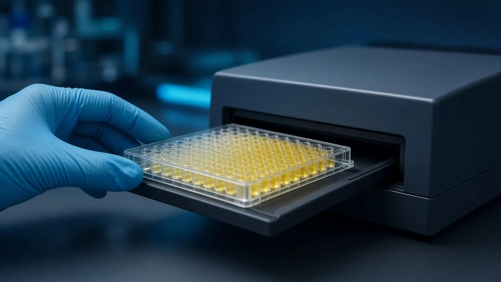
A single dilution of an antibody or a sample is a guess that happened to fit in the well. It can be too concentrated, so the blank climbs and every sample looks positive. It can be too dilute, so a real positive matches the blank and you change the biology instead of the pipette. The window you want is the range where specific signal rises as analyte rises and the blank stays flat. You only see that window by walking a series through it. Curves and formats are the setting, described in ELISA formats, controls and readout. This page is the titration habit that should have happened before the study plates were committed.
The symptom that means you never mapped the window
You are troubleshooting if one of these is true. Every well, including the zero, is dark, and diluting the sample does not separate them. Every well is pale, including a positive control that used to work, and adding minutes of substrate only lifts the blank. Or a datasheet dilution was copied onto a new matrix and the ranks of the samples reversed relative to an orthogonal measurement. Each of those can be a bad reagent. Each can also be a dilution that was never shown to sit in the window. Change one thing, and let the series tell you which axis is wrong.
Start by diluting the antibody you have, across a range wide enough that the ends look different from the middle. If the ends look the same, the reagent is dead or the plate is not seeing it, and the series has said so.
What too much and too little look like
Detection antibody in excess binds specific analyte and also sticks, weakly, to plastic, blocker and matrix. The blank rises. The difference between a low sample and the blank shrinks, and a standard curve compresses because the bottom asymptote has moved up. The temptation is to subtract the blank and continue. Subtraction of a large, noisy blank manufactures precision you do not have. Dilute the detection antibody until the blank falls and the mid standard still climbs. That pair of outcomes is the window.
Detection antibody too dilute leaves sandwiches unlabelled. The top of the curve sags, positives look low, and someone increases substrate time until the blank catches up. More substrate is the wrong knob if the enzyme was never bound. A series that includes a higher antibody concentration will show the top recovering while the blank is still quiet. If the blank rises before the top recovers, this antibody and this blocker do not have a usable window. Change the blocker or the clone. Do not split the difference at a single ugly dilution.
Sample dilution has its own shape. On the rising limb, diluting the sample dilutes the signal in proportion, and two dilutions agree after you multiply back. On the plateau, diluting the sample does not change the signal until you fall off the edge. Past a hook, diluting the sample increases the signal. A single neat well cannot show any of that. Two or three dilutions can. The same discipline is why a standard curve is a series and not a single calibrator point.
Typical steps are two-fold or three-fold, spanning at least a factor of ten to thirty around the datasheet suggestion so you are not sampling the same guess three times. The volumes and the incubation belong to the protocol of the reagent you opened. This is the layout logic, not a kit insert. Substrate timing notes from reagent makers, such as Promega protocols, describe development. Follow the bottle in your hand. Public plate designs on protocols.io are useful as maps of the idea. A measurement habit that states its dilutions openly is what a body such as NIST keeps pointing laboratories toward. It does not pick your titre.
Checkerboard is a class of experiment
A checkerboard, or a two-dimensional titration, varies two reagents. Capture dilution along one axis, detection dilution along the other, is the sandwich version. Primary against secondary is the indirect version. Each cell of the grid has a twin that lacks analyte, so you see signal and blank at every combination. You are not copying a plate map of microlitres from a manufacturer. You are choosing a small set of concentrations, often three or four per axis, that bracket the current guess.
The usable cell is not the darkest cell. The darkest cell is often excess reagent and a high blank. The usable cell is where the gap between plus-analyte and blank is wide, the blank is still near the substrate background, and neighbouring cells look similar enough that a small pipetting error will not kick you out of the window. A cell that works only at one razor-edge concentration will not survive the next operator.
If the whole grid is blank-high, the blocker or the wash is the problem and no antibody dilution will save it. If the whole grid is pale, including the highest concentrations, the enzyme, the substrate or the capture coat has failed. The grid is allowed to tell you to leave the antibody axis alone.
Hold the series to one plate when you can. A checkerboard split across two days has confounded dilution with day. The point of the grid is that the cells share a development.
Samples need the same honesty
Once the antibody window is chosen, the study samples still get a small series the first time you meet a new matrix. One dilution that lands on the steep limb, confirmed by a second dilution that agrees after correction, is a measured sample. One dilution taken on faith is the datasheet problem again, moved downstream. High samples hook. Low samples fall into the blank and should be reported as below the assay rather than as a precise small number. The curve shape that makes this obvious is the business of a bent standard curve. Use it.
| Pattern | High antibody | Low antibody | High sample | What it means |
|---|---|---|---|---|
| Blank dark, samples dark | Often the cause | Blank should fall if this was the cause | Blank stays dark even when sample is diluted | Dilute the antibody. A high sample is not the driver if the zero is already high |
| Blank quiet, samples pasted at the top | May be excess enzyme on a real sandwich | Top may sag if you cut too far | Can be the cause, including a hook if a dilution gets darker | Dilute the sample and read again. Do not lengthen substrate |
| Blank quiet, samples quiet, positive control quiet | Too low, or the reagent is dead | Same, more so | Irrelevant until a control works | Raise antibody once. If nothing moves, suspect coat, enzyme or substrate |
| Blank quiet, mid samples separated, dilutions agree | You are near the window | You may have left it | Samples are on the limb | Keep this region. Do not hunt for a darker well |
| One cell works, its neighbours do not | The window is too narrow to trust | Same | A pipetting accident is likely | Repeat the grid. Do not build a study on a singleton cell |
The series is only real if the pipetting is real
Every transfer in a serial dilution multiplies the previous error. A pipette that is short by a small fraction at each step walks the stated concentrations away from the liquid in the tube. The window you then "find" is a window in nominal units. Build the series with a calibrated pipette, a tip that fits, and the mixing the volume requires. Accurate micropipetting technique is the companion skill. Reverse pipetting of foamy antibody solutions, wiping a tip, and failing to pre-wet are enough to invent a titre. When two operators disagree on the usable cell, suspect the pipettes before you suspect the biology.
Independent check: make the chosen dilution two ways, once as a serial walk and once as a direct dilution from stock, and ask whether they match. If they do not, the series was arithmetic fiction. Repair it before any study sample is read against it.
Plate maps written after the read are how a dilution gets relabelled to match the hope. Write the map first. Include the blank twin of every antibody concentration. A series without blanks is a set of dark wells with no denominator.
Safety and the research limit
Antibody stocks may contain azide. Sample dilutions of serum, lysate or culture fluid carry the biosafety assignment of the parent material. Do the transfers in the containment that assignment names. The WHO Laboratory Biosafety Manual is a public reference for how institutions frame that choice. A carefully titrated research ELISA is still not a diagnostic test, and a dilution that lands on a curve is not a clinical reference interval.
Heat, evaporation and an open row of dilutions
An open dilution plate on a warm, dry bench loses water while you prepare the next reagent. Edge wells concentrate, the series tilts, and the usable cell moves toward whichever side evaporated. Cover the plate, work from capped tubes, and do not build the series beside a sunny window or under a vent. In humid weather a chilled plate collects condensation that dilutes wells you thought were sealed. Do not read a watery edge as a biological drop.
If a power cut stops the reader after a colorimetric series has been stopped, read it when power returns and note the delay. A series left half-finished overnight is two experiments. Start it again.
What to ask when you want reagents you can titrate
State the assay format, the matrix, the datasheet dilution you do not want to trust blindly, and the readout. Ask for the concentration of the reagent as supplied, so a series can be planned in actual units rather than in "fold" language that changes meaning when a vial is filled to a different volume. The reagents and chemicals catalogue is a route to related reagent classes. The molecular biology pathway places the assay among other methods. A titration plan can be discussed through the quote request. Ask the reply for the recommended starting range and the blocker class, and then run the series anyway. A single suggested dilution is the guess this page is about retiring.
Questions from the bench
Why is the dilution printed on a datasheet not already the answer?
The datasheet dilution was chosen on the supplier's antigen, plastic, blocker and reader. Your matrix and your conjugate shift the window. Treat the printed figure as the centre of a series you still have to run, not as a setting you may skip. A single well at that dilution cannot show you the neighbours that would have been cleaner.
What does a checkerboard add that a one-way series does not?
A one-way series varies one reagent and hopes the other is already right. A checkerboard varies two reagents at once, typically capture and detection, or primary and secondary, across a small grid. The usable cell is where specific signal has risen and the matching blank is still flat. A one-way series can miss that cell if the reagent you held constant was the one that needed to move.
How do I tell too much antibody from too much sample?
Too much detection antibody raises the blank and the sample together, so the difference between them shrinks. Too much sample can push a sandwich onto the plateau or into a hook, while the blank stays quiet. Dilute the reagent that changes the blank if the blank is the problem. Dilute the sample if the blank is quiet and the sample is pasted to the top of the reader.
Does a series rescue bad pipetting?
No. A serial dilution compounds every transfer error. The window you find is only as real as the volumes you delivered. A careful series with a poorly calibrated pipette is a careful map of the wrong concentrations. Fix the pipetting, then trust the pattern.
References
Manufacturer names identify published method classes. Trademarks remain with their owners. Catalogue records on this site are independent references for enquiry. They are not a statement of inventory, distribution rights or a supply commitment. This page is educational. It is not medical advice, a diagnostic protocol or a biosafety approval.
Catalogue
Related products and categories
These links follow the subject of the article into published manufacturer references. A listing is a reference for an enquiry, not a statement of stock or distribution rights.
Continue in this cluster
Related reading
ELISA formats controls and readoutELISA formats, controls and readout: how direct, sandwich and competitive assays turn a binding event into a number that stays inside the curve. Accurate micropipetting techniqueHow an air-displacement micropipette delivers a volume, how forward pipetting uses the two stops, and why temperature belongs in the result.Standard curves that bendWhy immunoassay standard curves are sigmoid, where the usable window sits, and what an early bend or a forced straight line does to a reported value.