
protocol overview
Hook effect and dilute-to-check
Why a very high analyte can make a sandwich ELISA look low, and how a planned dilution series separates a hook effect from a truly low sample.
- Author
- EVRINTH Editorial Team
- Published
- 8 October 2026
- Updated
- 8 October 2026
- Reading time
- 10 min
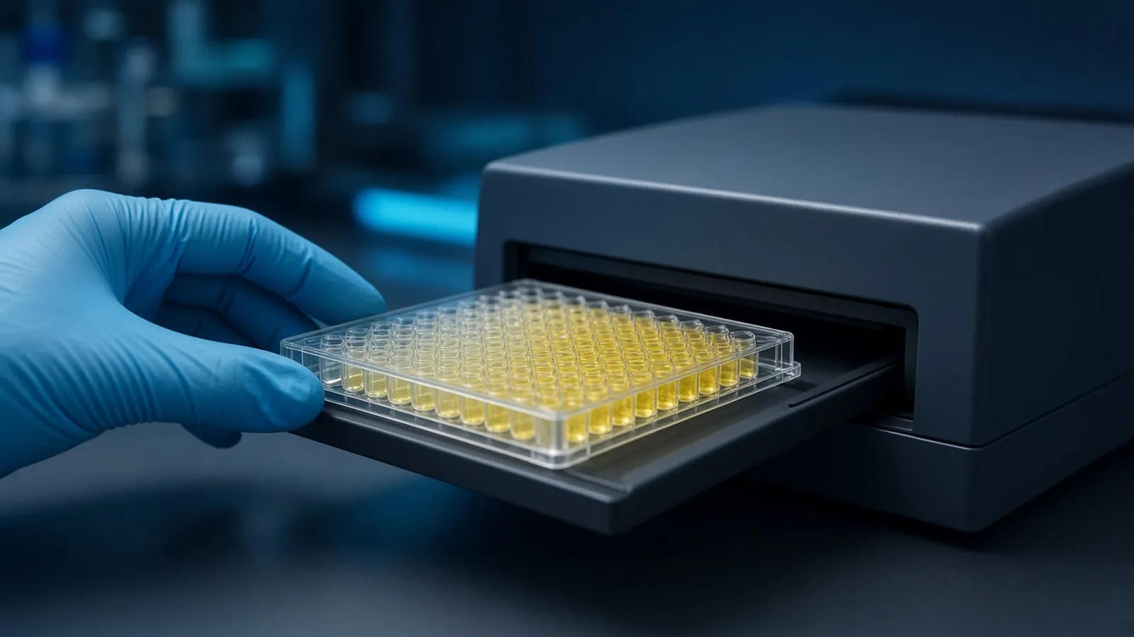
The hook effect is a sandwich ELISA lying by understatement. The analyte is not scarce. It is so abundant that capture antibody and detection antibody are each occupied by different analyte molecules, the sandwich between them is never completed, and the signal falls. The reader reports a modest number. A colleague who trusts that number will tell you the sample is low. The correction is dilution, done on purpose, before you believe the neat well. Formats and ordinary curves are described in ELISA formats, controls and readout. This page is the high-dose failure and the dilute-to-check that exposes it. It applies to research samples.
Who should plan the dilution before the plate is coated
Plan it when the analyte can span a wide range inside one experiment: an overexpression lysate next to an untransfected lysate, a stimulated culture supernatant next to a resting one, a recombinant prep, or a biological fluid you have not measured before. You do not know yet whether the darkest well is the highest analyte or the well that has already fallen off the far side of the curve. A single neat measurement cannot tell those stories apart.
The decision the check supports is narrow. Either the dilutions agree after you multiply back by the dilution factor, and you may report that concentration, or they do not, and you have no concentration yet. Averaging a hooked neat well with a valid dilution produces a third number that none of the wells contained.
How excess analyte dismantles a sandwich
A sandwich needs one analyte molecule to touch both antibodies. Capture antibody is stuck to the plastic, in limited amount. Detection antibody is in solution, also in limited amount. In the rising part of the curve, more analyte means more bridges and more signal.
Past a point, analyte is in excess of both partners. In a one-step incubation, free analyte binds capture sites and, separately, binds detection antibody in solution. Detection antibody that has already bound a free analyte molecule often cannot bind a captured one, because the epitope is occupied or the geometry is wrong. Captured analyte that has no free detection partner adds no enzyme. Signal therefore climbs, peaks, and falls as you add still more analyte. Plotted against concentration, the curve looks like a hook.
A two-step assay changes the order. Analyte binds the capture antibody first. Unbound analyte is washed away. Only then does detection antibody arrive, and it meets analyte that is already on the plate rather than a sea of free analyte. That removes the classic simultaneous hook. It does not grant an infinite top. Capture sites still saturate, so the curve still flattens, and a wash that leaves residual free analyte can reintroduce competition. Treat "two-step" as a reduction of risk, not as a proof that every neat well is inside the assay.
The primary antibody's affinity and the amount coated both move the concentration where the hook begins. A very tight pair hooks at a higher dose than a weak pair, but every limited-reagent sandwich has a dose it cannot swallow. Checking that the detection reagent really sees the analyte is a separate job, covered in choosing and checking a primary antibody. A hook is not nonspecific binding. Nonspecific binding paints the blank. A hook makes a high sample look like a moderate one while the blank stays quiet. Those two failures need different fixes.
What you actually plate
You need the sandwich reagents you already trust, a standard curve in a matched matrix, and enough of each research sample to run it neat and diluted. Diluent should be the matrix itself when the matrix is free of analyte, or the diluent the protocol specifies for that matrix. Diluting serum into plain buffer can change recovery and create a false disagreement that is matrix, not hook. Typical dilution steps are two-fold or three-fold. The right steps are the ones that walk a high sample back onto the steep limb. Follow the volumes and incubations the manufacturer states for the antibodies and the substrate. This overview is the logic, not a copied kit insert.
Include a zero matrix well so you can see background, and a mid-curve control you have run before so you know the plate itself developed. Public method sketches on protocols.io are useful for plate-map habits. The accession of the analyte you think is hooking can be checked on UniProt so you are not diluting the wrong protein's story.
The branch after the read
Read raw signal before you look at the software's concentration column. Then apply the rule.
If the neat well is light, and each dilution is lighter still, the sample is low or it sits on the rising limb. You may back-calculate any dilution that lands inside the standard curve, multiply by the dilution factor, and demand that those corrected values agree. If only the neat well is inside the curve and the dilutions fall into the blank, report the neat result and say the dilutions were below the assay.
If the neat well is light or moderate and a dilution is darker, stop calling the sample low. That rise is the signature of a hook, or of an inhibitor you diluted away. A further dilution that comes back down the far side of the curve, and whose corrected concentration agrees with the darker well, tells you which. Accept the agreeing pair. Discard the neat well for quantification. Record the pattern so the next person does not "clean up" the data by deleting the dark dilution as an outlier.
If every dilution is darker than the curve's top, you have not diluted enough. The sample is still on the plateau or still on the hook. Make a more aggressive dilution. Do not extrapolate above the top standard.
If dilutions disagree after correction even though all of them sit on the steep limb, you have a matrix or pipetting problem, not a clean hook. Do not average them into a compromise concentration.
| Neat signal | Diluted signal | Corrected concentrations | Interpretation |
|---|---|---|---|
| Low, and dilutions lower | Falls as expected | Neat value stands, dilutions may be below range | Consistent with a truly low or moderate sample |
| Moderate, dilution darker | Rises on dilution | Dilutions agree, neat is lower than they are | Hook effect. Report the dilutions, not the neat well |
| Above the top standard | Still above the top | None are inside the curve | Dilute further. Do not extrapolate |
| Inside the curve | Inside the curve but corrected values drift apart | No agreement | Matrix effect or a bad dilution series. Repeat before you report |
Other reasons a neat well looks falsely low
An inhibitor in the matrix can suppress enzyme or hide the epitope, and dilution can release the signal in a pattern that resembles a hook. The distinguishing follow-up is a spike. Add a known mid-curve amount of calibrator into the neat sample and into the dilution. Poor recovery only in the neat well means the matrix is quenching. A hook from analyte excess still recovers the spike once you are back on the limb, and the unspiked dilutions already agree with each other.
A dead detection antibody makes every well low, including the top standard. That is not a hook. Hooks are sample-specific. The standard curve on the same plate still climbs. If the standards are flat too, repair the reagents before you dilute the samples again.
Edge evaporation concentrates a well and can push it onto the hook or onto the plateau. If only the outer wells "hook", suspect the lid and the map, not the biology. A sample swapped with the blank is the boring version of a surprise. The plate map, written before the read, is the control for that.
Aerosols, serum and institutional biosafety
Diluting a research serum, a lysate or a culture fluid can make aerosols. Do it in the containment the biosafety officer assigned to that material, with the eye and hand protection that assignment names. Antibody reagents may contain azide. Substrate stop solutions are often acid. None of that changes because the scientific question is a hook. The WHO Laboratory Biosafety Manual is a public reference for how institutions set those rules. This dilute-to-check is a research control. It is not a procedure for diagnosing disease, and a low neat well must not be reported to a clinician as a low result.
Warm pipettes and a dilution that is not the one you wrote
A serial dilution on a warm, dry bench loses water from the open wells while you pipette the next row. The nominal two-fold step becomes something steeper, and two "agreeing" corrections agree only because both factors are wrong in the same direction. Cap tubes, work from a fresh diluent, and do not build the series in a plate sitting under a fan or in direct sun. If the room is hot enough that substrate development races, a hooked well and a valid high well can both hit the reader ceiling and look identical. Stop earlier and read, or you will call a hook a plateau.
Power cuts matter when the incubator holding a timed incubation stops and then restarts, or when you cannot read a kinetic plate. A stopped colorimetric plate can wait. Write down the clock time you added stop reagent so a delayed read is still tied to a finished reaction. A dilution series you have to repeat tomorrow is cheaper than a concentration you invented from a half-read plate.
What to ask when the format must survive a high sample
Tell the enquiry how high the analyte might go, whether you can run a two-step incubation, and whether the matrix is serum, lysate, medium or a purified prep. Name the antibody pair and the sample species. Ask whether the reagent design is simultaneous or sequential, because that choice moves the hook. The reagents and chemicals catalogue is where buffer and plate classes sit. The molecular biology pathway is the surrounding research context. A hook-aware layout can be discussed through the quote request. Ask for the incubation order in the reply. A measurement note from a body such as NIST will not list your analyte's hook point. That point is established on your plate, by dilution.
Dilute a sandwich sample until the dilutions agree
- 01Run the sample neat and at two or more dilutionsPlate the undiluted research sample beside at least two dilutions made in the same matrix or in the diluent the protocol names. Carry the standard curve on the same plate. Do not decide from the neat well alone.
- 02Plot signal against the dilution, not only against the back-calculated valueLook at the raw signal first. A real low sample stays low or falls further as you dilute it. A hooked sample rises into the steep part of the curve as analyte excess is relieved.
- 03Correct the dilution and demand agreementMultiply each in-range result by the dilution factor. Accept a concentration only where two dilutions, after that correction, agree within the spread you set before the plate was read.
- 04Reject the neat well when it disagrees with the dilutionsIf the neat well back-calculates lower than a diluted well, do not average them. Report the value from the agreeing dilutions, and record that the neat well was past the assay.
Questions from the bench
Why does a one-step sandwich hook more easily?
In a one-step, or simultaneous, sandwich the capture antibody, the analyte and the detection antibody meet in one incubation. At very high analyte, separate analyte molecules can fill the capture sites and the detection sites so the bridge between them never forms. A two-step assay washes unbound analyte away before detection antibody is added, which removes that particular route, though it does not make every high sample safe.
What should the signal do when a truly low sample is diluted?
It should stay low or fall. Dilution removes analyte, so a sample that was already on the rising limb or near the blank should not become darker. If a dilute well is darker than the neat well, treat the neat result as a hook until a further dilution explains it.
Does a two-step protocol abolish the hook effect?
It reduces the classic simultaneous hook. It does not define the problem out of existence. Capture antibody can still be saturated, the curve can still flatten, and a sloppy wash can leave enough free analyte to compete. Keep a dilution check for samples you expect may be extreme.
Is a hooked well evidence about a patient?
Not from this page. The pattern is a research observation about a sandwich immunoassay that was driven past its chemistry. Clinical assays that manage high-dose hook are validated for a stated specimen and a stated decision. Do not import a research dilution into a diagnostic claim.
References
Manufacturer names identify published method classes. Trademarks remain with their owners. Catalogue records on this site are independent references for enquiry. They are not a statement of inventory, distribution rights or a supply commitment. This page is educational. It is not medical advice, a diagnostic protocol or a biosafety approval.
Catalogue
Related products and categories
These links follow the subject of the article into published manufacturer references. A listing is a reference for an enquiry, not a statement of stock or distribution rights.
Continue in this cluster
Related reading
ELISA formats controls and readoutELISA formats, controls and readout: how direct, sandwich and competitive assays turn a binding event into a number that stays inside the curve. Choosing and checking a primary antibodyHow to choose and check a primary antibody: host, clonality, application claims, and controls that show the signal is the intended target.Dilution series instead of a single guessWhy one antibody or sample dilution is a guess, and how a dilution series or checkerboard finds the window where the blank stays flat.