
application
Immunohistochemistry is a different matrix
Why an antibody that works in an ELISA can fail on fixed tissue, and which controls make an immunohistochemistry result a research observation.
- Author
- EVRINTH Editorial Team
- Published
- 8 October 2026
- Updated
- 8 October 2026
- Reading time
- 8 min
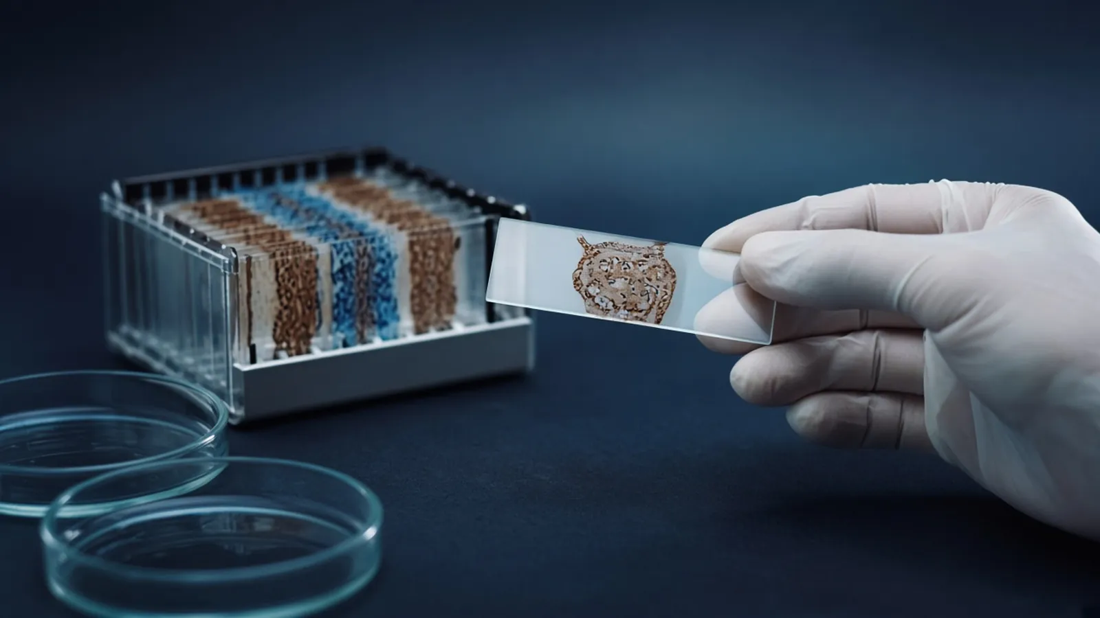
Immunohistochemistry asks an antibody to find its epitope inside a piece of tissue that has been fixed, embedded or frozen, cut, and stuck to glass. An ELISA asks an antibody to find its epitope in a well. The reagents can share a name and still be doing different chemistry. A dilution, a blocker and a development time that were honest on a plate are not a method on a section until the section says so. Plate logic stays in ELISA formats, controls and readout. Choosing the primary reagent is choosing and checking a primary antibody. The photograph on this page is a stained slide beside a rack of slides. The work below is research histology, not a pathology report.
What decision a tissue stain can support
You run immunohistochemistry when the question needs a place. Which cells in this research specimen carry the protein. Whether it sits in the nucleus, the cytoplasm or the membrane. Whether a treated group and a control group differ in that pattern. A well can tell you that an extract contained more signal. It cannot tell you that the signal moved from one compartment to another, because the extract has already destroyed the compartment.
The decision the stain supports is a comparison among sections that saw the same fixation, the same retrieval, the same antibody lot and the same development. It does not support a clinical diagnosis. It does not inherit validity from an ELISA datasheet.
Why the epitope the ELISA saw may no longer be there
Formaldehyde cross-links proteins. The cross-links preserve morphology and can bury or chemically alter the very stretch the antibody was raised against. An antibody that bound a native protein in a sandwich, or a denatured protein on a blot, can be silent on a formalin section, or it can bind only after a retrieval step you have not run. Alcohol and acetone fixation precipitate proteins and extract some of them. An antigen that survives formalin can wash out of an alcohol-fixed frozen section, and the reverse happens too.
The tissue is also full of other sticky surfaces: collagen, charged matrix, residual fixative, endogenous peroxidase or phosphatase, and immunoglobulin if the primary host matches the species of the tissue. A blocker that was enough for polystyrene can be irrelevant on a section, and a blocker that contains biotin can create a new stain in a tissue that is already biotin-rich. Transfer nothing unchanged. Not the dilution, not the blocker, not the minutes of development.
Antigen retrieval is a separate method with its own controls. The point here is only that you are not allowed to skip the fact of fixation. Look up where the protein is supposed to live, and in which compartments databases such as UniProt place it, before you interpret a surprising stain. The database is a prior. The slide can disagree for a real biological reason or because the antibody is on the wrong protein.
Controls that belong to the section
A secondary-only section, processed through every retrieval and detection step without primary antibody, is mandatory. Colour there is endogenous enzyme, biotin, or a secondary that sees the tissue. Quench endogenous peroxidase when the detection is peroxidase and the tissue is rich in it, such as blood-rich organs, and prove the quench on that control.
A negative tissue, or a negative cell pellet prepared as if it were tissue, should lack the target for a reason you can explain. A knockout animal or a cell line shown not to express the target is the strong version. A cell line is only as good as its identity. Misidentified lines are common enough that ICLAC exists to catalogue them, and culture collections such as ATCC culture guides document how reference lines are maintained. A negative section chosen only because it came out blank is not a control. It is a circular argument.
A positive tissue, if you have one with independent evidence that the protein is present, shows that today's retrieval and detection could have worked. A blank study section next to a blank positive control means the method failed. A blank study section next to a positive control that stained in the expected compartment is a result.
Run these on the same day as the study sections. A control slide from last month does not witness today's retrieval bath.
Localisation is part of the answer
Record where the colour sits. A transcription factor with a clean nuclear stain in the positive control and a nuclear stain in the sample is a coherent observation. The same antibody producing a cytoplasmic blush in every cell type, including the negative tissue, is nonspecific binding until proved otherwise. Edge staining of the section, stain trapped in folds, and stain that follows the knife mark are artefacts of processing. They are not expression gradients. Photograph the controls at the same exposure as the samples. A longer exposure on the sample alone manufactures a difference.
| Question | What ELISA evidence can say | What immunohistochemistry evidence can say |
|---|---|---|
| Is the analyte present in an extract? | Yes, if the curve and the blanks hold | No. A section is not an extract, and stain intensity is a poor quantity |
| Which cell type holds it? | No. The well has no anatomy | Yes, if morphology is intact and the controls are clean |
| Which compartment holds it? | No | Yes, and a compartment that contradicts the biology is a warning |
| Did fixation destroy the epitope? | The plate never asks | A negative stain beside a working positive control, or a rescue by retrieval, is how you see it |
| Is the antibody specific? | Only for the well format | Only for this fixation and this tissue. The ELISA result does not transfer |
Failure patterns that belong to slides
Uniform colour across every cell, including cells that should be negative, is a block or secondary problem. Repeat the secondary-only slide before you dilute the primary at random. Stain only at the edge of the tissue is drying or a thick edge, not a biological rim. Stain that appears after retrieval and vanishes without retrieval may be a true unmasking, or it may be retrieval damage that exposed sticky sites. The negative tissue has to pass through the same retrieval. If the negative tissue also wakes up, the retrieval opened the wrong thing.
A primary raised in the same species as the tissue, paired with a secondary against that species, stains endogenous immunoglobulin. The pattern follows plasma cells and serum, which can look temptingly specific. The secondary-only slide catches it if you are willing to look.
Do not quantify a brown precipitate with the same language you use for an ELISA curve. Stain intensity depends on section thickness, development time and haematoxylin counterstain. If you need a number, say what was scored, by whom, and against which controls. Do not import a four-parameter logistic.
Fixatives, solvents and institutional safety
Formaldehyde, xylene-class clearing solvents where a protocol uses them, and mounting media are chemical hazards with institutional rules. Tissue from humans or from infected animals carries a biosafety assignment that fixation may reduce and does not automatically erase. The WHO Laboratory Biosafety Manual is a public reference for that class of decision. Follow the local rule for how long a fixative must act before a section is handled at the bench you use. A research stain is still not a diagnostic report, however careful the solvent handling was.
Humidity, a drying rack and a warm afternoon
Sections that dry between antibody steps collect edge artefact first, and a humid room that later chills can drop water onto a dried slide and leave a tide mark. Cover the slides, keep them level, and do not interpret a tide mark as a gradient of expression. In a hot laboratory, peroxidase development runs faster. The development time you copied from a paper written in a cooler room can overshoot. Watch the positive control and stop when the compartment is clear and the negative tissue is still quiet, inside the window the detection protocol allows.
If a power cut stops a hybridisation oven or a retrieval steamer halfway, those slides are not a mild version of the method. Mark them and repeat. A partial retrieval is a different epitope exposure. Do not average it with slides that finished.
What to say when the antibody is for tissue
Tell the enquiry the fixation, the species of the tissue, the host species of any antibody you already have, and whether you need a method that avoids staining endogenous immunoglobulin. Say that an ELISA dilution is not the deliverable you want. Ask for the tissue and fixation in which the reagent was tried, and treat that statement as a starting point you will retest. The reagents and chemicals catalogue is a route to related reagent classes. The molecular biology pathway places the work among other research methods. An immunohistochemistry approach can be discussed through the quote request. Ask the reply to separate plate claims from section claims.
Questions from the bench
Why can a dilution that works in an ELISA fail on a tissue section?
The ELISA well holds antigen in solution or on plastic, usually at a concentration you chose. The section holds fixed cells, extracellular matrix and endogenous enzymes, and the epitope may be cross-linked or hidden. The antibody concentration that was optimal in the well is a guess on the slide. Titrate it again in the tissue.
What does a secondary-only slide tell you?
It tells you what the detection reagents, the block and the endogenous enzyme do with no primary antibody present. Colour on that slide is not target. If you skip it, you cannot tell a true stain from a biotin-rich tissue or from a secondary that sees immunoglobulin already in the section.
Why is the compartment of the stain part of the result?
A nuclear protein that appears only in the cytoplasm, or a membrane protein that fills the nucleus, is a finding you must explain before you call it expression. The explanation may be a real biological isoform, or it may be a cross-reactive antibody and a retrieval method that opened the wrong sites. Localisation is evidence, not decoration.
Can a research immunohistochemistry image be issued as a pathology report?
No. Diagnostic histopathology is a regulated medical activity with its own validation, controls and reporting duties. A research section supports a research statement about the specimens you stained and the controls you ran. Keep those boundaries in the figure legend.
References
Manufacturer names identify published method classes. Trademarks remain with their owners. Catalogue records on this site are independent references for enquiry. They are not a statement of inventory, distribution rights or a supply commitment. This page is educational. It is not medical advice, a diagnostic protocol or a biosafety approval.
Catalogue
Related products and categories
These links follow the subject of the article into published manufacturer references. A listing is a reference for an enquiry, not a statement of stock or distribution rights.
Continue in this cluster
Related reading
 ELISA formats controls and readoutELISA formats, controls and readout: how direct, sandwich and competitive assays turn a binding event into a number that stays inside the curve.Choosing and checking a primary antibodyHow to choose and check a primary antibody: host, clonality, application claims, and controls that show the signal is the intended target.Fixation and antigen retrieval conceptsHow formaldehyde and alcohol fixation change epitopes, and how to test one retrieval class against morphology before you lock a method.