
comparison
Quantifying DNA with a spectrophotometer
A spectrophotometer number is a concentration only after a blank and a pathlength. Compare a pedestal with a cuvette before you trust the reading.
- Author
- EVRINTH Editorial Team
- Published
- 8 October 2026
- Updated
- 8 October 2026
- Reading time
- 8 min
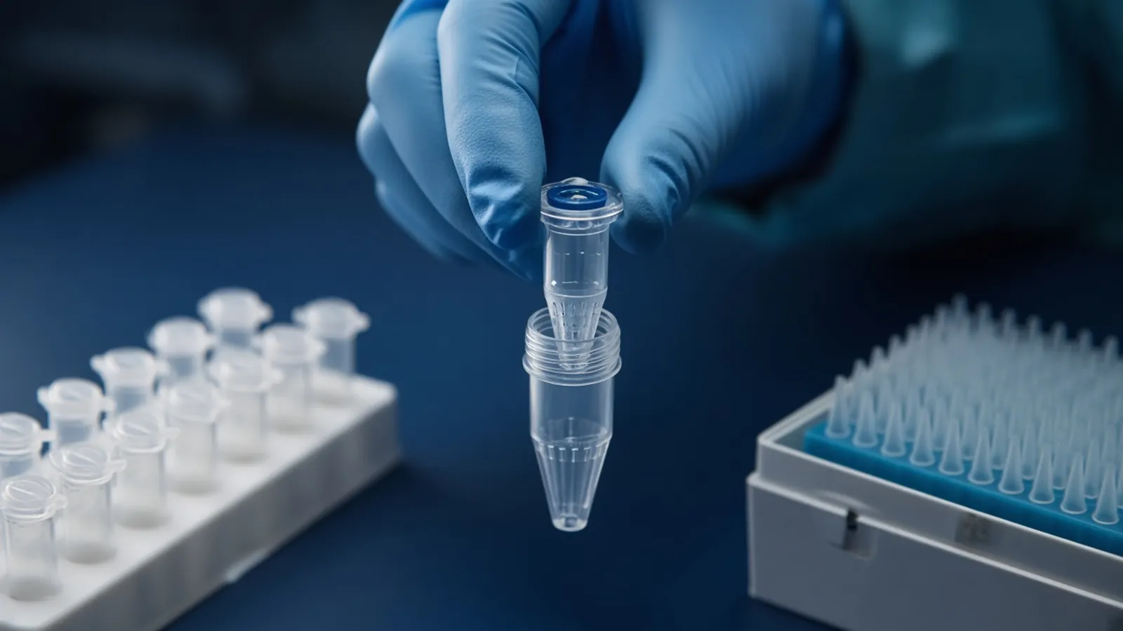
Quantifying DNA with a spectrophotometer is a decision about whether a displayed number is allowed to enter the next calculation. The instrument measures how much ultraviolet light the solution stops. You turn that measurement into a concentration only after two conditions are true: the blank is the buffer the sample sits in, and the pathlength used in the calculation is the pathlength the light actually travelled. A number without a blank and a pathlength is not a concentration. This comparison is for research benches choosing between a microvolume pedestal and a cuvette. It does not certify an instrument and it is not a clinical result.
Sample lysis and a silica column, a bead wash, or a precipitation step decided what else is in the tube. Those method classes are compared in how DNA extraction methods differ. Quantification starts after that choice, when you need a mass for a digest, a ligation, or a library. Write the blank identity, the pathlength the software assumed, and the conversion factor beside every number.
Beer-Lambert, the blank, and the pathlength
The Beer-Lambert relation says absorbance rises in proportion to how strongly the molecule absorbs, how concentrated it is, and how far the light travels through the liquid. In symbols, absorbance equals the extinction coefficient times concentration times pathlength. Rearranged, concentration equals absorbance divided by extinction and pathlength. Shorten the path and the same solution gives a smaller absorbance. Forget to scale back to the path you intended, and you report a concentration that is too low.
The blank is the zero. You place the elution buffer, with no sample, in the light path and tell the instrument that this absorbance is the baseline. Everything the sample then shows is the extra absorbance above that buffer. Skip the blank and the buffer, the dust, and the dirty window become part of the sample. Blank with the wrong liquid and you subtract a baseline the sample does not have.
Pathlength is the distance the beam travels inside the liquid, not the length of the instrument. A classical cuvette is often a 1 cm path. A microvolume pedestal holds a column of liquid that may be a fraction of a millimetre, then software scales the raw absorbance to a 1 cm equivalent if someone set that option. Those are different measurements that can be made to agree. They do not agree by default.
Water quality belongs in this step when the blank is water or a buffer you made yourself. Organic traces from a tired purification cartridge absorb in the ultraviolet and move the baseline. The grades and the failures are set out in laboratory water types and where they fail. Blank with the finished buffer, not with a jug that merely looks clear.
The usual 50, 40 and 33 conversions
For a 1 cm path, laboratories commonly convert one absorbance unit at 260 nm with three planning factors. About 50 ng per microlitre for double-stranded DNA. About 40 ng per microlitre for RNA. About 33 ng per microlitre for single-stranded DNA. These factors assume a fairly average base composition and a solution whose absorbance is mostly nucleic acid. Oligonucleotides are better converted with a sequence-specific extinction coefficient when the sequence is known. A plasmid and a PCR product can share the factor of 50 and still differ in how much of the absorbance is really long DNA.
The instrument pathlength setting has to match the factor. Apply 50 to a raw short-path absorbance and the concentration collapses. Apply 50 a second time to a value the software already scaled to 1 cm and the concentration doubles. Read the menu. If the display already says nanograms per microlitre, find which factor it used before you multiply again.
Ratios sit beside the conversion and do not replace it. A260/A280 near 1.8 for DNA and near 2.0 for RNA hints at protein, not integrity or identity. A260/A230 often falls when chaotrope, phenol, or carbohydrate remains. Free nucleotides still absorb at 260 nm and inflate the mass. RNA integrity is a separate question from either ratio.
Pedestal and cuvette are different instrument classes
A microvolume pedestal sandwiches about one or two microlitres between two optical surfaces. The path is short, and the software may offer a 1 cm-normalised absorbance. That suits a silica-column elution you cannot spend entirely on the measurement. A bubble, a dust speck, or a salty ring is a large fraction of a one-microlitre droplet. Very dilute samples produce a tiny raw absorbance, so the scaled number jumps. Wipe as the instrument note describes, then blank again. A pedestal that still holds the previous sample will quantify that leftover.
A cuvette gives a stated path, commonly 1 cm, sometimes a shorter path that is printed on the cell. You need more volume, often hundreds of microlitres depending on the cell, so it is a poor way to spend a tiny elution. It is a fair way to measure a dilute sample when you can spare the volume, because the longer path produces a larger absorbance above the noise. The blank belongs in a matched cell or in the same cell, rinsed into the buffer you are using. Fingerprints on the window, a scratched face, and a cuvette left wet with the previous buffer all shift the reading. If the method asks for a 1 cm factor and the cell is a 0.2 cm path, the arithmetic has to say so. The cell does not know which factor you memorised.
| Choice | Microvolume pedestal | Cuvette |
|---|---|---|
| Volume | Often one to two microlitres | Hundreds of microlitres, set by the cell |
| Pathlength | Short, then scaled in software if that option is on | Stated on the cell, often 1 cm |
| Dilute samples | Small raw signal, noisy scaled number | Longer path gives more signal when volume allows |
| Typical miss | Dirty surface, evaporation, wrong factor | Scratched window, mismatched blank, ignored path |
| After a small elution | Leaves most of the tube for the assay | Can consume the tube to measure it |
Two pedestals from the same family still disagree if one was blanked and the other was not.
A workflow with branch points
Mix the eluate. A silica-column tube can stratify if it sat. Blank with the same bottle of elution buffer you used.
On a pedestal, confirm the blank sits near zero from about 220 to 320 nm, then load the sample and check the pathlength and the factor. If a repeat droplet wanders, wipe, blank, and read again. Do not average a wild set into false precision. In a cuvette, rinse with the blank, confirm the path printed on that cell, and dilute in the same buffer if the absorbance is clipped. A clipped trace is an overrun, not a high concentration.
Branch on the shape of the spectrum. Nucleic acid rises toward 260 nm. A shift toward 270 nm can be phenol. A high shoulder at 230 nm often tracks leftover chaotrope. Keep that absorbance out of a sensitive enzyme calculation until a cleanup or a dye assay says how much is really DNA. Return to the extraction method rather than tuning the factor until the number looks convenient.
Failure modes worth separating
A zero or negative concentration after a visible pellet usually means the blank was dirtier than the sample, or the pedestal never made contact with the droplet. Clean, reblank, and reseat the liquid. Do not conclude the prep failed on that single screen.
A very high number from a clear, tiny elution can be real, or it can be a factor applied twice. Check the arithmetic before you dilute the tube. Carry-over on a shared pedestal is the other quiet error: the previous plasmid is still on the surface, so every sample looks rich. Clean until a buffer blank is flat. If an extraction blank absorbs like the samples, the reader is reporting the method, not the specimen.
Free nucleotides and RNA absorb at 260 nm, so a double-stranded mass can look larger than a later dye assay will agree. When the next step needs that mass, confirm with a selective method.
Safety and the limit of the claim
The ultraviolet lamp stays enclosed in normal use. The specimen may still be infectious, and chaotropic salt or phenol is a hazard of the prep. Follow the safety data for the bottle you opened. Institutional biosafety rules decide whether that eluate may sit on an open bench. The WHO laboratory biosafety manual is background for that local decision. A research concentration is a planning figure for the next enzyme reaction, not a diagnostic or forensic measurement.
Warm rooms and a blank that drifts
In a hot laboratory a one-microlitre droplet shrinks while a dialogue box is still open. Water leaves, the DNA stays, and the concentration rises. Read promptly. A power cut can reboot a reader into a default factor you did not choose, so blank again after the lamp is warm. Humidity does not rewrite Beer-Lambert. It does make a salty residue stick to the lower pedestal.
What to put in an enquiry
State the nucleic acid, the elution volume you must keep, whether the path will be a pedestal or a cuvette, and the assay that will consume the mass. Name the blank buffer. Use the sample preparation catalogue and the sample preparation pathway, and send the requirement with the quote request. The nucleic acid isolation enquiry reference is a place for specimen and assay questions. Ask whether a quotation is possible. These pages are enquiry references, not a statement that a particular reader is already in a laboratory.
Questions from the bench
Can I multiply a raw absorbance by 50 and call it a DNA concentration?
Only when that absorbance is already expressed for a 1 cm path and the molecule is double-stranded DNA in a clean buffer. A short-path pedestal reports a small raw absorbance, and the software may or may not have scaled it. Confirm the pathlength setting and the factor before you write nanograms per microlitre in the notebook.
What should the blank be?
Blank with the buffer the DNA is actually in, measured in the same vessel class as the sample. A water blank of a Tris-EDTA eluate treats the buffer itself as nucleic acid and shifts the baseline, especially near 230 nm. If the blank will not settle, clean the pedestal or the cuvette and prepare a fresh aliquot of that buffer.
Why do two readers disagree on the same tube?
They may be applying different pathlength corrections, different factors, or different blanks. A dirty pedestal and a scratched cuvette do not share an error. Re-read both against the same elution buffer and write down which factor each instrument used.
Does a clean 260 reading prove the silica column prep will ligate?
No. Absorbance estimates how much material absorbs at 260 nm. It does not show fragment length, and it can miss inhibitors that do not absorb there. The extraction class still has to match the assay, as discussed in the comparison of DNA extraction methods.
References
Manufacturer names identify published method classes. Trademarks remain with their owners. Catalogue records on this site are independent references for enquiry. They are not a statement of inventory, distribution rights or a supply commitment. This page is educational. It is not medical advice, a diagnostic protocol or a biosafety approval.
Catalogue
Related products and categories
These links follow the subject of the article into published manufacturer references. A listing is a reference for an enquiry, not a statement of stock or distribution rights.
Continue in this cluster
Related reading
How DNA extraction methods differHow silica columns, beads, precipitation and organic extraction change DNA yield, length and inhibitors, and which class fits the assay you actually plan toA glossary of purification termsDefine chaotrope, bind-wash-elute, yield, purity ratios and RIN, then walk a purification so those words describe one real workflow.Blood saliva and tissue as starting materialExplain how EDTA blood, heparin blood, saliva and solid tissue behave as extraction inputs, and why a serum tube is not whole blood.