
troubleshooting
Salting-out and organic extraction concepts
Reason from a dirty phenol interphase or a low 260/230 back to salting-out or organic phase chemistry, without treating solvents as a casual recipe.
- Author
- EVRINTH Editorial Team
- Published
- 8 October 2026
- Updated
- 8 October 2026
- Reading time
- 10 min
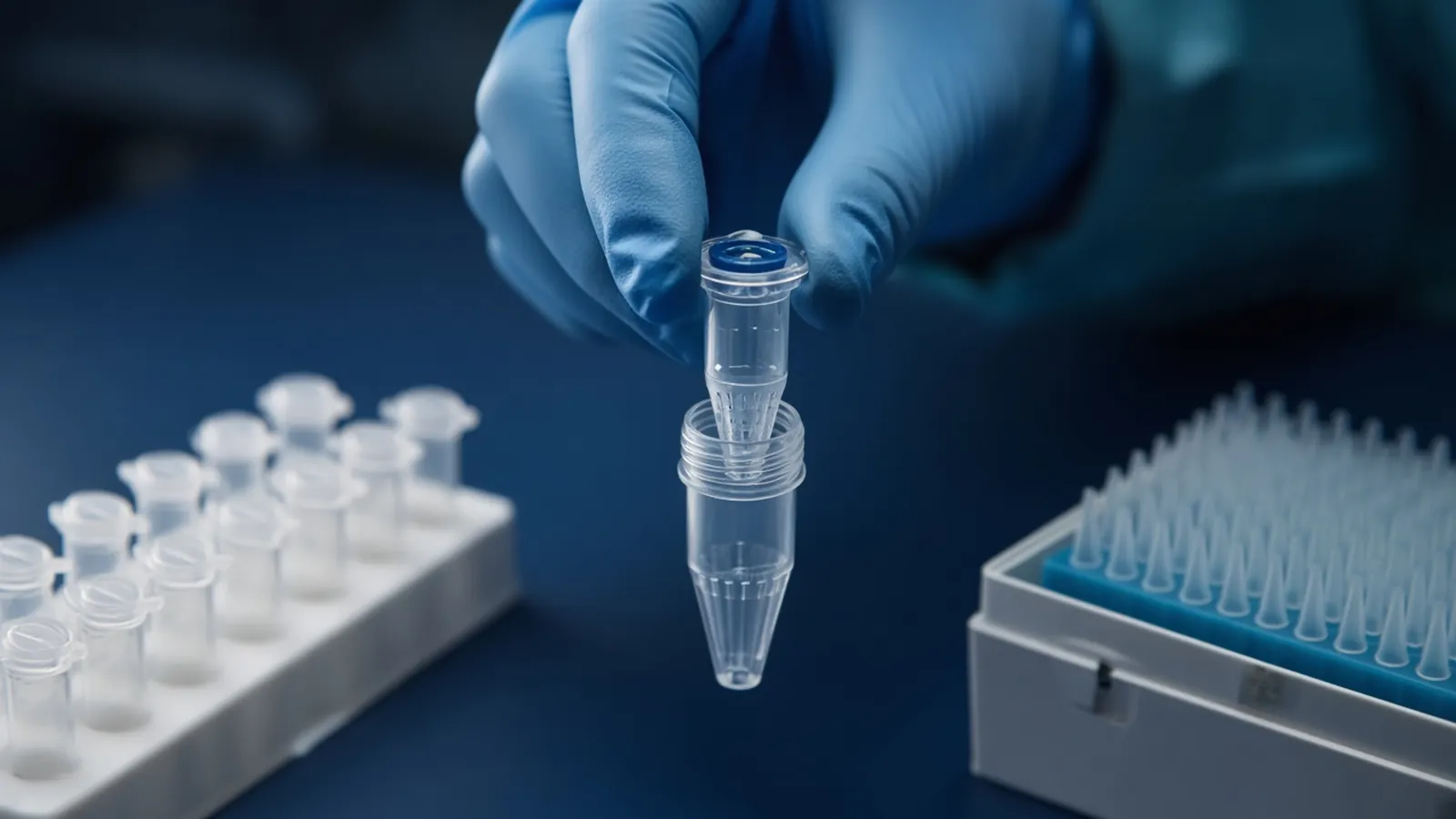
Salting-out and organic extraction are two older ways to persuade protein to leave nucleic acid alone. They still solve real problems, and they still produce two familiar troubles: a dirty interphase you pipetted through, and a low 260/230 that appears after the tube looks "done". This page is a way to reason about those symptoms. It is not a pipetting recipe, and it is not an invitation to handle phenol or chloroform unless your institution has already made that chemical decision.
Both methods sit beside silica columns and beads in how DNA extraction methods differ. If a column already gives you the length and the cleanliness the assay needs, you do not graduate to solvents for status. RNA handled with phenol has extra rules, covered from the integrity side in protecting RNA during extraction.
What each chemistry is trying to partition
Organic extraction uses phenol to denature proteins. Chloroform, often together with a small amount of an antifoam alcohol in classical mixtures, sharpens the boundary and reduces how much phenol lingers in the aqueous layer. After centrifugation you can see an aqueous phase, an organic phase, and an interphase of denatured protein. Nucleic acid stays in the aqueous phase only when the pH matches the protocol. DNA protocols keep that phase from being strongly acidic, so DNA remains aqueous while protein goes to the phenol and the interphase. Acidic phenol conditions used for RNA move DNA the other way. Taking the wrong phase, or using the wrong pH, is a lost sample rather than a low yield you can wash back.
The interphase is not a third product to harvest. It is the trash layer. The closer the pipette comes to it, the more protein and the more phenol travel with the aqueous phase. A thick interphase means there was a lot of protein or the tube was overloaded. Leaving a generous margin of aqueous phase behind sacrifices yield and protects the prep. That trade is the whole practical art. Volumes, shake times and spin settings belong in the institutional SOP and on the bottle, not in a remembered sketch.
Salting-out skips the organic phase. After a detergent lysis, a high concentration of salt drives proteins out of solution. A spin pellets that protein. Nucleic acid remains in the supernatant and is then usually precipitated with alcohol, washed, and dissolved. The salt identity and the concentration are part of a named protocol, not a pinch. Done gently, this route can keep genomic DNA long, because you never asked a solvent interface to do the separation and you need not vortex the final pellet. The leftovers are different: residual salt, and polysaccharides that co-precipitate with the alcohol step. Those leftovers are exactly what a low 260/230 is good at hinting.
A silica column can appear at the end of either story as a cleanup, if the SOP says the aqueous phase or the dissolved pellet may be bound and washed. The column removes some salt and some small inhibitors. It does not repair DNA you scooped from the interphase along with a plug of protein, and it does not make phenol safe to pour.
Symptom, then the reasoning path
Start by naming which method you actually ran. A low ratio means different things after phenol than after salt.
If the interphase is thick, the sample was too rich in protein for the volume of the SOP, or the tissue was not digested enough before the solvent. The reasoning path is yield versus contamination. Take a smaller fraction of the aqueous phase from a repeat, or reduce the input, rather than plunging the tip to the white band to "get it all". If proteinase digestion was optional in that SOP and you skipped it, the thick layer is the skipped step showing itself. Do not invent a new solvent ratio to compensate.
If there is no clean boundary, you have an emulsion. Warm rooms and vigorous shaking both encourage it. The reasoning path is to stop adding energy. An SOP may permit a longer spin to let droplets coalesce. It may permit a specific accepted trick. If it permits neither, the tube is a question for the method owner, not a prompt to pour more chloroform because a forum said so. An emulsion that you pipette anyway becomes a dirty aqueous phase with a fine 260 reading and a dead enzyme later.
If the aqueous phase was cloudy after you transferred it, you took the interphase with you. Expect a low 260/280, a gummy pellet, and a restriction enzyme or a polymerase that sulks. The reasoning path is a further cleanup that your SOP already allows, such as a repeated phase separation under that SOP or a silica wash of the aqueous phase. Re-measuring the cloudy liquid on a spectrophotometer does not extract the protein.
If 260/230 is low after an organic prep and the blank is truly the elution buffer, suspect phenol carry-over or carbohydrate. Phenol also distorts the region near 270 nm, so 260/280 may look oddly high or oddly low rather than "a bit protein". Enzymes that die, and a chemical smell that should not be in an aqueous eluate, support solvent carry-over. The reasoning path is a cleanup class, not a second casual extraction written from memory. If the assay still works and the ratio is the only complaint, write the limitation down. A ratio is a hint. A dead ligase is a result.
If 260/230 is low after salting-out, suspect the salt you added on purpose, or polysaccharide from the tissue, or EDTA in the resuspension buffer. A clean alcohol wash, as that precipitation protocol already describes, is the first reasoning step. A silica column is the second if salt survives the wash and the assay cares. Do not conclude the protein precipitation "failed" solely from 260/230. Protein shows more honestly at 260/280 and on a gel that will not digest.
If both ratios look poor and a blank of unused buffer looks poor too, the spectrophotometer is in the story. Clean the surface and blank again before you condemn the phase separation. Dirty blanks move these numbers even when the prep was textbook.
RNA integrity does not fall out of either ratio. A phenol RNA prep can show a plausible 260/280 and still be degraded because the tissue waited. Judge RNA on a trace. Acidic phenol that accidentally went into a DNA prep will make the DNA vanish from the aqueous phase while any RNA looks, briefly, like a success. Read the pH class on the bottle before you troubleshoot yield.
| What you see | Likely class of cause | Reasoning step that stays inside an SOP |
|---|---|---|
| Thick white interphase | Protein overload or poor digestion | Leave a margin of aqueous phase; reduce input next time |
| Milky tube, no boundary | Emulsion from over-mixing | Stop shaking; only the SOP may extend the spin |
| Cloudy aqueous phase | Interphase was pipetted | Cleanup the SOP allows; do not call it pure |
| Low 260/230 after phenol | Phenol or carbohydrate | Cleanup class; connect it to enzyme failure if enzymes die |
| Low 260/230 after salting-out | Residual salt or polysaccharide | Alcohol wash class, then a column only if the SOP agrees |
| Low 260/230 and a dirty blank | The reader, not the prep | Reblank with the real buffer before repeating the extraction |
| Good ratios, short DNA on a gel | Shear during mixing | Length is a handling problem, not a phase problem |
| No DNA after acidic phenol | Wrong pH class for DNA | Confirm the phenol protocol was a DNA protocol |
What not to do with a disappointing tube
Do not shake harder, and do not pour an unwritten extra volume of phenol because the interface looked ugly. Residual solvent is an inhibitor and a chemical exposure. A silica column cleanup belongs only where the SOP already sent the aqueous phase. A low 260/230 is not a moral score: after salting-out it can be the salt you added and then failed to wash, and a high value still does not prove RNA integrity or a working PCR. Pair the ratio with enzyme failure, a brown pellet, or a gel that shows shear. If the same SOP is clean on a cell pellet and dirty on tissue, reduce the tissue input to the amount that SOP was built for. Do not scale solvent by eye.
Solvents are an institutional hazard decision
Phenol burns skin, and it is readily absorbed. Chloroform is toxic. Both need a cabinet, eye and skin protection, and a waste stream the institution has already approved. Salting-out avoids that solvent pair and still uses alcohol, which is flammable, and high salt, which is irritating. Chaotropes appear only if you move to a silica column afterwards. None of this page is a substitute for the safety data on the bottle you opened.
Specimens from people or from infected animals stay under the biosafety decision your committee has made. The WHO laboratory biosafety manual and the WHO biosafety health topic are public background. They do not authorise a containment level, and they do not turn an organic extraction into a clinical test. A research DNA prep is not a diagnostic result.
If your laboratory is not set up for phenol waste, the troubleshooting path for a dirty interphase is to stop using that method, not to do it more carefully in a shared sink. Salting-out or a column-and-bead class may already meet the assay. That is a legitimate endpoint of troubleshooting.
Writing the hazard into the request, not into a corridor bottle
The operational failure in many shared buildings is a solvent bottle that arrived because someone ordered "DNA extraction reagent" and received phenol without a cabinet to match. When you enquire, say whether phenol and chloroform are permitted in your laboratory at all. If they are not, ask for a salting-out class or a silica class and say so in the first sentence. If they are, name the waste path you already have, the specimen, the fragment length, and whether the target is DNA or RNA so the pH class cannot be swapped by accident.
Use the sample preparation catalogue and the sample preparation pathway to point at the non-solvent alternatives when those are the ones you are allowed to run. Send the constraint with the quote request. The nucleic acid isolation enquiry reference is a place to put the scientific question. It is an enquiry reference. It does not mean an organic extraction is performed for you. Ask whether a quotation is possible. The acceptance check is a clean phase boundary or a salt-washed pellet, a believable blank, and an assay that still works, not a ratio achieved by any means.
Questions from the bench
Why is there a white layer between the two liquid phases?
That interphase is denatured protein and other insoluble debris parked between the aqueous layer and the organic layer. A thin line is ordinary. A thick, cloudy band means the sample was protein-rich or overloaded. If your pipette touched it, protein is now in the nucleic acid, and a pretty later column will not change the fact that the aqueous phase was already dirty.
The tube looks milky and will not form two layers. What should I conclude?
You have an emulsion, which is the two phases mixed into droplets. More shaking usually makes the droplets smaller and the wait longer. Stop. The institutional SOP may allow a longer centrifugation to sharpen the boundary. Improvising extra solvent volumes at the bench is how a hazard becomes a personal recipe. If the SOP does not cover the emulsion, ask the person who owns the method before you add anything.
A low 260/230 after salting-out means the salt failed, does it not?
It means something that absorbs near 230 nm is still there. Residual salt from the precipitation, carbohydrate from the tissue, or a dirty blank can all do that. The protein-precipitation step can still have worked. Wash the nucleic acid the way that precipitation protocol already specifies, or move the dissolved pellet onto a silica column if your SOP allows, and blank the spectrophotometer with the buffer you actually used.
Can I switch a DNA phenol protocol to RNA by using the same bottle colder?
Cold is not the switch. pH is. DNA stays in the aqueous phase when that phase is kept in the neutral to slightly alkaline range used for DNA protocols. Acidic phenol mixtures drive DNA out of the aqueous phase and are the RNA-style separation. Using the wrong pH loses the molecule you wanted. RNA integrity is then a separate question of how fast RNases were stopped, not of how cold the phenol felt.
References
Manufacturer names identify published method classes. Trademarks remain with their owners. Catalogue records on this site are independent references for enquiry. They are not a statement of inventory, distribution rights or a supply commitment. This page is educational. It is not medical advice, a diagnostic protocol or a biosafety approval.
Catalogue
Related products and categories
These links follow the subject of the article into published manufacturer references. A listing is a reference for an enquiry, not a statement of stock or distribution rights.
Continue in this cluster
Related reading
How DNA extraction methods differHow silica columns, beads, precipitation and organic extraction change DNA yield, length and inhibitors, and which class fits the assay you actually plan toA glossary of purification termsDefine chaotrope, bind-wash-elute, yield, purity ratios and RIN, then walk a purification so those words describe one real workflow.Blood saliva and tissue as starting materialExplain how EDTA blood, heparin blood, saliva and solid tissue behave as extraction inputs, and why a serum tube is not whole blood.