
glossary
Troubleshooting a smeared protein gel
How to branch a smeared protein gel: overload, salt, DNA, degradation, polymerisation, buffer, bubbles, detergent and a leaking well.
- Author
- EVRINTH Editorial Team
- Published
- 8 October 2026
- Updated
- 8 October 2026
- Reading time
- 7 min
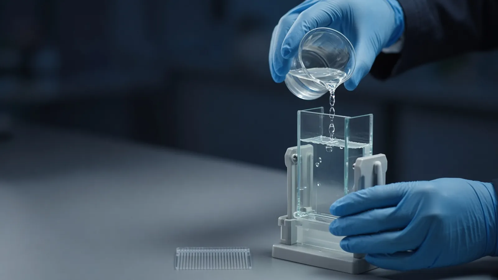
A smear on a protein gel is a shape you can name. It is not yet a diagnosis. Overloaded wells, salt, viscous DNA, a degraded sample, a poorly polymerised gel, exhausted initiator, the wrong running buffer, bubbles, excess detergent and a hot sample that leaked before the voltage came on all stretch bands into streaks. The useful move is to branch. This page is a research troubleshooting guide. It does not replace the casting recipe you already validated.
What a fair lane is allowed to prove is set out in reading a protein gel. How much protein you thought you loaded depends on the assay in how laboratories estimate protein concentration and on the lysate-specific comparison in comparing quantification assays for a lysate.
Look at the marker before you blame the sample
The ladder is the control that splits the causes into two families. If the marker is also smeared, smiling, or stalled, the gel, the buffer or the run is the lead. Sample stories about proteases and inclusion bodies can wait. If the marker is sharp and one lane is a curtain, that lane's contents or how it was loaded are the lead. If every sample smears and the marker does not, they share a preparation habit: the same lysate buffer, the same boil, the same overloaded volume.
Write down the acrylamide percentage, whether the gel is Tris-glycine or a Bis-Tris system with its own buffer, and whether the sample was reduced. A smear in the wrong buffer system is a mismatch, not a mysterious protein. Reused running buffer, diluted by mistake or exhausted after too many runs, produces fuzzy dye fronts and poor stacking. Replace it before you recast the whole box of plates.
Sample branches when the ladder is honest
Overload is the most common research smear. Too much protein in the well exceeds the stacking capacity. The band becomes a vertical streak, often darker at the top, and neighbouring lanes can distort. The cure is a smaller load, chosen from a real quantification, not from "the tube looked concentrated". A five-fold dilution series on the next gel tells you whether the protein was always a smear or only a smear at that load.
Salt does a quieter version of the same damage. High salt in the sample slows stacking. Bands leave the well late, smile, or spread. Elutions from ion exchange, or lysates in high-NaCl buffers, do this even when the protein amount is modest. Dilute into sample buffer, or desalt, rather than stuffing the well to compensate.
DNA makes a lysate viscous. The sample strings out of the tip, sits in the well, and drags protein down the lane as a streak from the top. A nuclease treatment, or a harder clarifying spin of the lysate before you ever make the gel sample, is the class of fix. Shearing with a needle is a habit some labs use. It is also a way to lose material on plastic. Prefer the method your lysate protocol already names.
A degraded sample smears downward and loses the high-mass bands you saw in an earlier aliquot of the same prep. Proteases remain active in SDS until the sample is fully denatured, and they work, more slowly, in the cold. Compare a freshly lysed aliquot, kept with inhibitors and loaded promptly, with the tube that waited. If only the old tube smears, degradation is the lead. If both smear, look back at overload and DNA.
Too much detergent, beyond the SDS already designed into the sample buffer, fuzzes bands and can produce micellar junk near the dye front. Lysis buffers that were loaded almost neat, plus a second dose of SDS sample buffer, are a typical route. Dilute the lysate. Detergent that will later matter for mass spectrometry is a separate problem, discussed in detergents that ruin a later mass spec step.
Gel and loading branches when the ladder suffers too
Incomplete polymerisation leaves a soft, wavy, or grainy gel. Bands wiggle even when the protein is pure. Ammonium persulfate is the radical source. TEMED catalyses release of those radicals. Both are concept-level consumables here: they age, APS solutions especially once made up, and a warm humid room shortens the life your recipe assumed. If the gel feels tacky, casts with a wavy interface, or runs as a smear including the marker, cast again with initiator you still trust. Follow the recipe's proportions. This page will not invent a microlitre table.
Air bubbles under the comb or against the plates force current around a gap. The lane bends or splits. If you see a bubble, pull the comb, refill if the gel is still liquid, or abandon that well. Do not interpret a bent band around a bubble as a doublet.
A hot sample leaks. Boiled tubes are fluid, and a well that is loaded and then left on the bench allows protein to diffuse sideways before the run stacks it. Leaks between wells from a torn well wall do the same. Load cooled sample, start the voltage promptly, and discard torn wells. A streak that begins between lanes, rather than inside one well, is a leak until proved otherwise.
| What you see | First branch | Leave for later |
|---|---|---|
| Marker and samples all smeared | Buffer identity, buffer age, polymerisation | Protease story |
| Marker sharp, one lane a curtain from the well | Overload, DNA viscosity, that sample's salt | Recasting the plates |
| High-mass bands gone, smear to the dye | Degradation versus a fresh aliquot | A new acrylamide percentage |
| Wavy interface, soft gel | APS and TEMED age, incomplete set | The biological sample |
| Band bent around a gap | Bubble or torn well | The sequence of the protein |
| Neighbouring lanes share a streak | Leak of a hot or overfilled well | Inclusion-body naming |
How to change one variable
Repeat the gel with a single change. Dilute the suspect lane, or replace the running buffer, or recast with fresh initiator. Changing percentage, load, buffer and sample history in one afternoon produces a second smear you still cannot assign. Keep an aliquot of the original sample so the comparison is real.
A lane that smears only above a certain mass, with sharp bands below, can be a genuine mixture of oligomers or a glycosylated population. That pattern is data if it survives a lower load and a fresh gel. Call it a mixture only after the artefact branches have been tried.
Safety and what a smear is not allowed to decide
Acrylamide monomer is a hazard until the gel has polymerised, and even then the laboratory's waste rules apply. This guide does not authorise a diagnostic electrophoresis method. A smear is not an identity, not a purity percentage, and not evidence that an inclusion body failed to refold. Those claims need a designed comparison.
Heat, humidity, and a run that stops
Warm rooms speed the death of APS solutions and soften gels that were left to set on a sunny bench. Humidity drips into open plates and dilutes the top of a gel that was meant to be a stacking layer. Cast, overlay, and comb within the time your recipe expects, and keep the initiator bottle closed.
A power cut mid-run lets bands diffuse. Restarting an hour later does not restore the stack. Record the interruption and rerun from the remaining sample rather than measuring migration on a gel that sat without current. Ice under the tank is a local habit for very long runs. It does not repair a smeared load.
What to ask for when the gel itself is the problem
If you are sourcing acrylamide mixes, buffers, stains or markers, describe the symptom: marker smeared or sample smeared, gel chemistry, and whether the gel set firmly. Those reagent classes are in the reagents and chemicals catalogue. A smeared expression check is also a reason to state, in the protein expression enquiry reference, whether you need a soluble fraction or a washed insoluble fraction, and what a readable gel must show. Put the symptom in the quote request with the gel percentage and the buffer system, so the reply matches the branch you are actually on.
Questions from the bench
If the marker is sharp and the sample is smeared, where do I look?
The gel and the running buffer have already shown they can resolve a clean standard. Look at load, salt, DNA, detergent and whether that particular sample was degraded or leaked. Changing the acrylamide percentage first wastes the clue the marker just gave you.
Does a downward smear always mean proteolysis?
A smear toward the dye front is compatible with cleavage, and also with overload, residual salt and a sample that never stacked. Proteolysis becomes the lead when high-mass bands disappear, the lane looks digested compared with a fresh aliquot, and inhibitors or colder handling change the pattern.
Why mention old APS and TEMED if I should follow the gel recipe I already have?
Ammonium persulfate starts the polymerisation and TEMED accelerates it. Both are consumables that decay, and a soft or wavy gel is what that decay looks like. The point is to treat initiator age as a branch, then cast again with reagents your recipe still considers active.
Can I interpret a smeared lane as an inclusion body?
An inclusion body is a claim about an insoluble expression fraction, not about blur. A smeared pellet lane can be overload or DNA. Wash and load a defined amount, beside a supernatant lane, before you name the aggregate.
References
Manufacturer names identify published method classes. Trademarks remain with their owners. Catalogue records on this site are independent references for enquiry. They are not a statement of inventory, distribution rights or a supply commitment. This page is educational. It is not medical advice, a diagnostic protocol or a biosafety approval.
Catalogue
Related products and categories
These links follow the subject of the article into published manufacturer references. A listing is a reference for an enquiry, not a statement of stock or distribution rights.
Continue in this cluster
Related reading
Reading a protein gelHow to read a protein gel: what SDS-PAGE bands, smears, ladders and loading differences can support, and what they cannot identify. How laboratories estimate protein concentrationHow laboratories estimate protein concentration with absorbance, dye-binding and copper assays, and why the number depends on the standard.Comparing quantification assays for a lysateBradford, BCA and absorbance at 280 nm on a dirty lysate: which interference wins, why two kits disagree, and which standard to name.