
glossary
Troubleshooting flat amplification curves
How to read a flat qPCR trace: empty wells, dead reverse transcriptase, inhibition, seal failure and the wrong channel are different failures.
- Author
- EVRINTH Editorial Team
- Published
- 8 October 2026
- Updated
- 8 October 2026
- Reading time
- 9 min
The amplification plot stayed flat, and the software wrote undetermined where a quantification cycle should have been. That drawing is a result, and it is almost never one result. An empty well, a dead reverse transcriptase, a channel the instrument was not asked to read, and a genuine absence of template can all look alike until you branch. This glossary names the trace states and the forks between them. How a working relative assay is supposed to behave is in RT-qPCR for relative expression. The place of that assay in a wider study is in from cells to a gene expression result.
Flat is a fork
Start with the question the plate was meant to answer. A flat sample well is evidence about the transcript only if three other things are true: the positive control rose, the no-template control stayed quiet, and the raw optical trace, not only the analysed plot, shows that this well was read in the right channel. Skip any one of those and "not expressed" is a story you have not earned.
Reporting habits for real-time experiments are set out in the MIQE guidelines. A flat well still needs a reason in the notebook. Undetermined is not a biological category.
Fluorescence is not the transcript
A real-time instrument watches fluorescence against cycle number. A dye or a probe becomes bright as product accumulates. The software estimates a baseline from early cycles, subtracts it, sets a threshold, and calls the crossing point. The familiar sigmoid is that corrected curve.
Several words get used for drawings that are not the same object.
The amplification plot is the corrected view. It is what most people screenshot. It can hide a real rise if the baseline window was wrong.
The multicomponent view, sometimes called the raw view, shows dye channels before that correction. Passive reference dyes, target dyes and background are separated here. This is the view that tells you whether the camera saw anything.
Plateau is the late flat top of a successful curve, after reagents or instrument range are exhausted. A plateau after a proper rise is normal. A line that is flat from cycle one is not a plateau. An early plateau, where the curve is already high and flat in the baseline window, is a third state: too much template, a saturated dye, or a correction that started too late.
No amplification means the corrected and the raw target channel both stay flat. Something the chemistry needed was absent or inactive, or the product never became fluorescent in the channel you recorded.
Mixes, plates and channels
The reaction class is a master mix matched to the detection chemistry: an intercalating dye, or a probe with its own fluorophore. The instrument method has to acquire that fluorophore. A probe written as a green channel while the plate was run as a different reporter will be flat on the plot you are staring at, and alive on the channel you did not open.
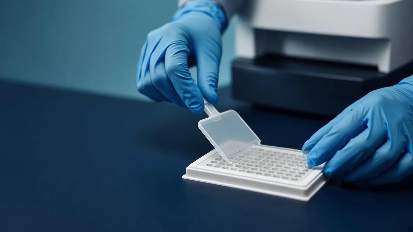
Plasticware is part of the optics. A white qPCR plate and an optical film exist so the light path works. A seal meant for ordinary endpoint PCR is not automatically an optical seal. The photograph of this workflow is hands seating film with an applicator. A film that is lifted at a corner lets water leave. Volume falls, salts rise, and the late cycles misbehave or the signal never forms.
Reverse transcriptase is a separate enzyme from the polymerase that copies DNA during the qPCR. A dead reverse transcription kills RNA assays and leaves a DNA control, if you included one, able to amplify. Forgotten primer is a setup error: the mix is complete except for the oligonucleotide that defines the product. An empty well is a pipetting miss. Those are different forks, and they are distinguished by which wells failed, not by the adjective flat.
Primers still have to be the right primers. A pair that cannot bind has the same drawing as a pair you forgot. NCBI Primer-BLAST is where you check what a pair can bind. It will not tell you whether this well received them.
Reading the plot, then the raw view
Take the wells in a fixed order.
Open the amplification plot for the channel the assay was supposed to use. Note which wells are sigmoid, which are flat from the start, and which rise immediately and sit in plateau. Note whether the flat wells cluster by sample, by row of a multichannel pipette, or by the edge of the plate.
Then open the multicomponent or raw view for one flat well and for one sigmoid well. If the flat well's target dye rises in the raw view, the baseline or the threshold ate a real curve. Revisit the baseline window. Abundant template that amplifies during the baseline cycles is the usual reason. If the raw target dye is also flat, there is no product fluorescence to rescue with settings.
Check the passive reference if the mix uses one. A reference dye that is missing because of a pipetting miss, or saturated, can warp the corrected curve even when product exists. The raw view shows the reference separately from the target.
Only then interpret biology. A sample that is raw-flat, beside a positive control that is sigmoid, on the correct channel, is a sample or reverse-transcription problem. It is not yet a claim that the gene is off.
Curve states worth naming
| What you see | Raw view | The branch it supports |
|---|---|---|
| Flat from cycle one, positive control sigmoid | Target dye flat | This well or this reverse transcription, not the whole mix |
| Flat, and the positive control is flat | Target dye flat on both | Primer, mix, channel, cycler or a plate-wide seal failure |
| Corrected plot flat, raw dye rising | Target dye rises | Baseline window or threshold, not a dead well |
| Already high, then flat | Dye high from the first cycles | Too much template or a saturated signal. Do not call it negative |
| Edge wells flat, centre wells fine | Often erratic | Seal or evaporation. Repeat before any biological claim |
| No-template well rises | Dye rises in the control | Contamination or primer artefact. Sample flats are not interpretable as clean negatives |
Inhibition can flatten a curve or shove it so late that the run ends first. A late, struggling curve is a different glossary entry from a line that never leaves baseline. How to separate inhibition from low template is the neighbouring decision, and it should not be collapsed into the word flat.
NTC, sample, and positive control
The no-template control contains the mix without the nucleic acid you claim to be testing. If it is flat and the positive control rises, the reagents can work and they are not already full of product. If the no-template control rises, you have contamination or a primer artefact. A flat sample beside a rising no-template control is not a clean negative. It is an uninterpretable plate. Contamination control, as a habit, is the subject of PCR controls and contamination control.
If samples and the no-template control are flat and a DNA positive control rises, suspect the reverse transcription or the RNA, not the qPCR polymerase. A previously successful cDNA from the same primers is a useful extra well: if that cDNA works today and today's reverse transcription does not, the enzyme or the RNA input is the fork.
If every well is flat, including controls, stop looking at genes. Confirm the run finished, confirm the channel, confirm primers were added, and confirm the film actually sealed. A multichannel skip can empty a row and leave the rest alive. That pattern is pipetting, discussed as a skill in accurate micropipetting technique.
Reference genes do not rescue a flat target by being present on a spreadsheet. If the reference is also flat in that well, the well failed. If the reference rises and the target is raw-flat, you may be looking at a low or absent target, and you still report the limits of the assay rather than a precise fold change of zero.
Causes that share one drawing
Dead reverse transcriptase, forgotten primer, empty well, wrong channel, seal failure and true absence share a screenshot and almost nothing else. The way through is the pattern across wells plus the raw optical trace. Sequencing is not the next step for a flat plate. If the question was transcriptome-wide, RNA-seq has its own library failures. It will not explain a qPCR well that never received primer. Method notes collected by Addgene are a reminder that controls are part of the written protocol, not a mood.
Not a diagnostic trace
A research qPCR trace does not approve a diagnostic result. Biosafety for the sample, especially RNA from human or infected material, is an institutional decision. Optical instruments and hot blocks are ordinary electrical and heat hazards. Follow the safety notes for the dyes and mixes you use. This glossary does not assign a clinical meaning to an undetermined well.
Seals, humidity and a run that stopped
Optical film that is applied in a humid room, or handled with damp gloves straight from a freezer, seals poorly. Edge lifting shows up after the block heats. If a pattern of failures sits on the rim, reseal a repeat and do not interpret the first file. In a building that drops power, a run that stops mid-cycle is an aborted programme. The partial curves are not quantification cycles. Restart only after you know whether the block finished, and treat an interrupted plate as failed unless a control strategy you wrote in advance says otherwise.
Master mixes follow the storage on their label. A warm shipping delay or a freezer that thawed is a reason to test a known positive before trusting a plate of scarce cDNA. "Room temperature" setup in a hot laboratory is not the temperature a protocol from a cooler climate assumed.
What to include if you ask for help with an assay
Describe the channel, whether the positive control and the no-template control were flat or not, and whether the raw view agreed with the corrected plot. Say whether the samples were RNA through a reverse transcription or already cDNA. Those facts decide the fork. A discussion of targeted assays can sit beside the mRNA sequencing enquiry reference when the scientific question may belong in sequencing instead, and beside the differential expression analysis enquiry reference when the end product is a comparison rather than a single plate. Neither page means a run is already under way.
Reagents and plates are classes in the molecular biology catalogue. The assay's place in sample-to-result work is the nucleic acid analysis pathway. Put the trace pattern, not a screenshot alone, in the quote request.
Questions from the bench
Does a flat amplification line mean the gene is absent?
Not by itself. A flat line means the software did not see fluorescence cross the threshold in that channel. The well may be empty, the primer may be missing, the reverse transcription may have failed, or the dye may be on a channel you did not acquire. Absence of transcript is only one of those branches, and only after the controls have worked.
Why would the amplification plot be flat when the raw view still rises?
The amplification plot is usually baseline-corrected. If the true signal rose during the cycles used as baseline, subtraction can flatten a real curve. The multicomponent or raw view shows the dyes before that correction. A rise there means the analysis settings ate the curve. A flat raw trace means no product fluorescence was recorded.
The positive control is flat as well. Where should I look first?
Look at the reaction and the instrument, not at the biology of every sample. Forgotten primers, a dead mix, the wrong channel, a block that did not run, or a seal that failed across the plate all hit the control too. Samples become interpretable again only after a control can amplify.
Can a poor seal look like a negative sample?
Yes. Evaporation changes volume and salt, and an optical seal that is not seated can kill the signal, especially in edge wells. The pattern is often spatial, at the rim of the plate, rather than grouped by biological condition. Treat a rim pattern as a plate failure until a resealed repeat says otherwise.
References
Manufacturer names identify published method classes. Trademarks remain with their owners. Catalogue records on this site are independent references for enquiry. They are not a statement of inventory, distribution rights or a supply commitment. This page is educational. It is not medical advice, a diagnostic protocol or a biosafety approval.
Catalogue
Related products and categories
These links follow the subject of the article into published manufacturer references. A listing is a reference for an enquiry, not a statement of stock or distribution rights.
Continue in this cluster
Related reading
 From cells to a gene expression resultHow a laboratory goes from cells to a gene expression result, and how RNA quality decides between a focused RT-qPCR assay and RNA-seq.A glossary of expression termsPlain definitions of Cq, TPM, counts, FDR, UMIs and the other expression words that cause arguments when they are used loosely.A heatmap is a picture not a conclusionHow clustering, colour scale and row scaling change a gene-expression heatmap, and why the differential table and the contrast remain the result.