
troubleshooting
Loading controls are for blots not every gel
Actin, GAPDH and tubulin support a blot loading argument. A purification gel is judged by the band pattern and the total protein you loaded.
- Author
- EVRINTH Editorial Team
- Published
- 8 October 2026
- Updated
- 8 October 2026
- Reading time
- 6 min
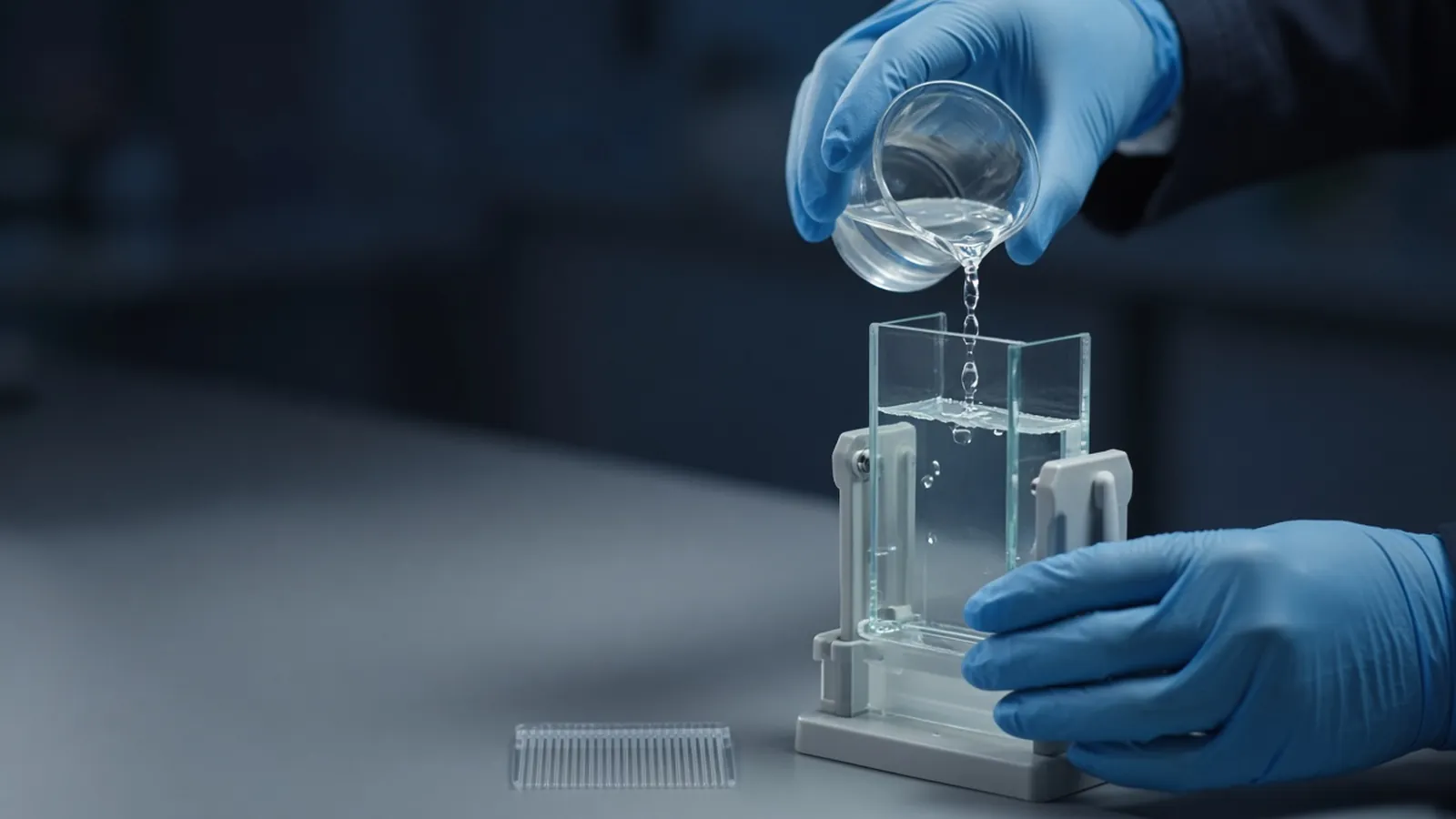
Actin, GAPDH and tubulin became famous on membranes. They are a poor habit to copy onto every Coomassie gel of a purification. A loading control, in the blot sense, is an argument that lanes received similar amounts of cellular protein and that transfer did not favour one lane. A stained purification gel is an argument about which bands are present and how intense they are relative to the mass you loaded. Mixing those arguments is how a methods section acquires a control that cannot do the job. The transfer itself is described in western blot from gel to membrane. How to read bands without over-claiming them is in reading a protein gel.
What a blot control is allowed to say
On a western blot of lysates, the primary detection is often one scarce polypeptide. You cannot see the rest of the proteome in that image. A second probe for a protein that you believe is stable across the comparison stands in for "these lanes started equal". Actin, GAPDH and tubulin are common choices because they are abundant. Abundance is also their weakness. The band saturates. A treatment that truly changes the housekeeping protein makes the control a second experiment, not a ruler.
The control also absorbs transfer failure. If one lane transferred poorly, both the target and a honest loading control should fade. If the target fades and the control does not, you have a clue that the loss is not simply a empty lane. If both fade, you do not get to claim down-regulation. None of this logic requires the control to appear on a Coomassie gel of four chromatography fractions. Those fractions are not cells, and the stain already shows total protein.
A purification gel is a pattern, not a housekeeping assay
Judge a purification by the load and the pattern. Early fractions should show many bands if the load is high enough to see contaminants. The pool you call purified should show the species you intended, at the mass you expect under the reducing conditions you used, with contaminants described rather than ignored. Equal volumes of fractions with different concentrations are not equal loads. Quantify, or load a defined fraction of each pool. The assay behind that microgram number matters, and it is the subject of how laboratories estimate protein concentration.
People sometimes ask for an actin blot of a bacterial lysate to "prove equal loading" of a recombinant protein. Escherichia coli is not a mammalian cytoskeleton. A mammalian loading control has nothing to bind. The right comparison is total protein loaded, or a stain of the same gel. If the recombinant band grows after induction while the background pattern stays similar, that is the induction argument. It does not need a GAPDH caption copied from a eukaryotic paper.
Saturation and the total-protein alternative
A saturated control lies politely. Film, a chemiluminescent image pushed too far, or a Coomassie band past the linear stain range all clip the signal. Two lanes look equally dark because both are at the ceiling. Load less, shorten the exposure, or use a detection range you have checked with a dilution series of one lysate. The dilution series is the control for the control.
A total-protein stain on the membrane, such as a reversible stain used before blocking, supports a different sentence: the lanes contain similar amounts of transferred protein overall. It does not claim that any one gene product was constant. That is often the better sentence when the treatment is severe enough to change metabolism, because GAPDH is a metabolic enzyme and actin is not immune to real biology. Stain-free or other total-protein detection systems make the same class of claim when they are used within their range. They are not decoration under a saturated target band.
| Surface | Equal signal supports | It does not support |
|---|---|---|
| Housekeeping blot | Similar load and transfer of that one protein, if the band is not saturated and the protein is truly stable | Purity, identity of the target, or equal loading on a Coomassie purification gel |
| Total-protein membrane stain | Similar transferred protein across many bands | That a specific housekeeping gene was unchanged |
| Coomassie purification gel | The visible pattern at the stated total-protein load | A cellular loading control, or molar purity when bands overlap |
| Target band alone | Nothing about loading | Down-regulation, yield, or purity |
When the control itself moves
Treat a changing loading control as a result. If GAPDH falls with the treatment, normalising the target to GAPDH can manufacture a fold-change. Show the control, and consider a total-protein normalisation if the scientific question is "per unit protein". If actin differs because the lanes were pipetted unevenly, fix the load with a quantified rerun rather than stretching the image.
On a purification, the failure mode is different. A single dark band can hide a co-migrating contaminant of the same mass. Loading less reveals whether the band splits. Loading more reveals faint contaminants. Both loads belong in the notebook. A saturated Coomassie band of the product, with the rest of the lane invisible because the scan was cropped, is not a purity figure. HUPO and related proteomics practice are a reminder that identity is a measurement, not a position on a gel.
Troubleshooting in order
Start with the load bookkeeping. Were the lanes equal micrograms, equal volumes, or equal fractions of a culture? Those are three different experiments. Then ask whether the control is in range. Then ask whether the control protein is biologically allowed to be constant in this design. Only then interpret the target. If a purification gel is ugly, quantify again and rerun matched loads before you add an antibody against a housekeeping protein that the sample may not contain.
A power cut during a run, or samples that sat warm while a blot tank waited, can degrade lanes unequally. A loading control will not repair proteolysis that already differed between tubes. Note the interruption, and do not average those lanes with a clean repeat as if they were the same handling.
Limits and an enquiry
Research blots and research gels are not diagnostic electrophoresis. Biosafety of the cells or the lysate is decided by your institution. Antibodies and stains have their own handling notes.
When you enquire about blot or gel reagents, say whether you need a lysate comparison or a purification series, which load you intend to match, and whether a total-protein stain or a single-protein control carries the claim. Reagents and chemicals is the relevant catalogue category. A quote should carry the detection class and the sample type. Questions about how a purified protein will be documented can be raised against the custom protein expression and purification enquiry reference. That page supports a discussion of the method. It is not evidence that a purification service is being operated.
Questions from the bench
Should every Coomassie gel include actin or GAPDH?
No. Those proteins are loading arguments for immunoblots of complex cellular samples. A purification gel is judged by how the band pattern changes across fractions and by how much total protein you loaded. An actin antibody on a nickel elution does not rescue a badly quantified lane.
Why do equal control bands still mislead?
A control that is saturated, on the blot or in the image, stays dark across a wide range of loads. The lanes can differ and the control looks flat. Stay inside the linear range of the detection method, or the equal-band claim is only a picture of saturation.
Is a total-protein stain the same as a housekeeping blot?
It is a different claim. A membrane stain or a stain-free total-protein signal reports the protein that transferred, across many bands. A single housekeeping polypeptide reports that one protein, which may itself have changed with the treatment. Use the claim that matches the sentence you want to write.
What should I report for a purification gel instead?
Report the load as micrograms of total protein from a named assay, or as a fraction of the pool, and show the marker and the fraction identity. Describe extra bands. Purity is a statement about that pattern, not about actin.
References
Manufacturer names identify published method classes. Trademarks remain with their owners. Catalogue records on this site are independent references for enquiry. They are not a statement of inventory, distribution rights or a supply commitment. This page is educational. It is not medical advice, a diagnostic protocol or a biosafety approval.
Catalogue
Related products and categories
These links follow the subject of the article into published manufacturer references. A listing is a reference for an enquiry, not a statement of stock or distribution rights.
Continue in this cluster
Related reading
Reading a protein gelHow to read a protein gel: what SDS-PAGE bands, smears, ladders and loading differences can support, and what they cannot identify.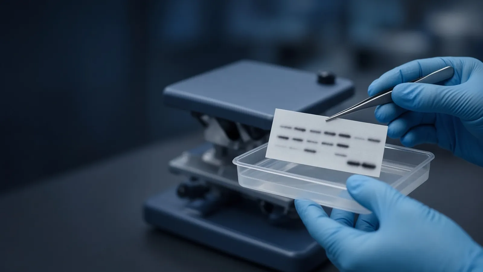 Western blot from gel to membraneHow a protein moves from an SDS gel onto a membrane, how antibodies create a band, and which controls separate transfer failure from a real signal.
 How laboratories estimate protein concentrationHow laboratories estimate protein concentration with absorbance, dye-binding and copper assays, and why the number depends on the standard.