
selection guide
Removing PCR inhibitors from difficult samples
Pick dilution, a further cleanup, or an inhibitor-tolerant polymerase when heme, humic acid, heparin or leftover solvent stops a PCR.
- Author
- EVRINTH Editorial Team
- Published
- 8 October 2026
- Updated
- 8 October 2026
- Reading time
- 9 min
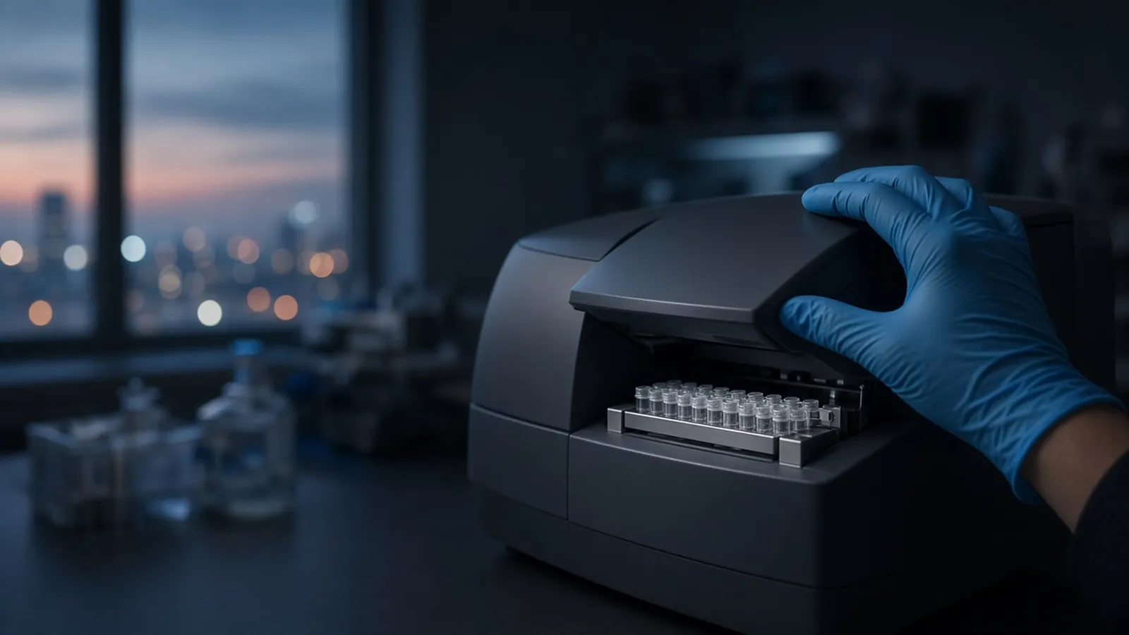
The photograph on this page is a gloved hand closing a thermal cycler on a strip of tubes. The inhibitor is discovered in that block, when a well stays dark or a lane stays empty. It is not removed by the lid. Removal, dilution, or a more tolerant enzyme happens before the strip is loaded. This guide is how to choose among those three, for research PCR from a difficult matrix. It is not a clinical test and not a kit insert.
Controls that make the choice interpretable are set out in PCR controls and contamination control. The extraction that either carried the inhibitor or washed it away is compared in how DNA extraction methods differ.
What is actually stopping the enzyme
Polymerases fail for chemical reasons that differ by specimen. Naming the likely molecule is how you avoid a random second kit.
Heme and haemoglobin come from blood, especially when red cells are lysed in a crude prep. They inhibit many common polymerases. Heparin comes from heparin collection tubes and can follow DNA through casual purification. EDTA from an EDTA blood tube is a different problem: it chelates magnesium the enzyme needs, and it usually yields to dilution or to a modest input into a magnesium-balanced mix. Do not treat every anticoagulant as heparin.
Humic and fulvic acids come from soil, sediment, compost and some environmental swabs. They colour extracts brown and inhibit PCR at levels a spectrophotometer may not flag as "protein". Polysaccharides come from plants, stool and some bacteria. They make lysates viscous and they inhibit or they simply entangle the template. Bile salts come from faecal samples and from some microbiome matrices. They are small, inhibitory, and easy to carry if the wash was timid.
Phenol comes from organic extraction that was pipetted too close to the interphase. Ethanol comes from a column or bead wash that was not removed. Guanidine and other chaotropes come from sample lysis buffers when the wash step was skipped, shortened, or overwhelmed by overload. All three are method leftovers rather than specimen biology. They tell you the purification was unfinished. A brown plant eluate may still be biology. A tube that smells of solvent is the method.
A260/A280 does not sort this list. A ratio near 1.8 can belong to a heparinised extract that will not amplify. RNA integrity, if you are about to reverse-transcribe, likewise says the strands are long enough and says nothing about bile or ethanol in the same droplet. Sample lysis can release the inhibitor (heme from a red cell, polyphenols from a leaf) at the same moment it releases the target. A harsher lysis is not automatically a cleaner PCR.
Three responses, and the criteria that pick one
Dilution is the fastest response. Inhibitors that act by mass often fall below their effect before the target does, if the target was abundant. A 1-in-10 dilution that turns a negative into a positive, or that rescues a spike, has diagnosed inhibition or overload. You can stop there when the positive is all the research question needed. You cannot stop there when you needed a quantitative claim, because you also diluted the template, or when the diluted target disappears. Dilution costs nothing but sensitivity.
A further cleanup is the response when the target is too scarce to dilute and the inhibitor is something a bind-wash or a precipitation can leave behind. Ethanol, guanidine, salt and much phenol are the usual wins for a fresh silica or bead wash. Heparin, humic acids and polysaccharides are less obedient. They sometimes need a chemistry aimed at them: a size-selective precipitation, a PVP-class additive already in a plant or soil method, or a resin class sold to strip inhibitors. Follow that chemistry's protocol. Do not chain three unnamed kits because the first failed. Each step spends yield and can introduce a new leftover.
An inhibitor-tolerant polymerase class is the response when this PCR is the end of the experiment, dilution would drop a rare target, and a full cleanup is losing the DNA. Some enzyme formulations are built to accept blood, soil or humic matrices that stop a classical Taq-type mix. They are a class with a published tolerance, not a universal solvent. Follow that enzyme's buffer and its template limit. They do not pull heparin out of the DNA. If a later ordinary enzyme must see the same tube, clean it anyway. Tolerance is for the well in the cycler, not a permanent property of the eluate.
Use this order of questions. Did a clean positive control amplify on this run? If it did not, you do not have an inhibitor story. You have a mix, a primer, or a cycler story. Did a spike inside the sample fail while the same spike in water worked? Then the sample matrix is inhibitory. Is the target known to be abundant in this specimen type? Dilute first. Is the target scarce, and is the inhibitor a wash leftover such as ethanol or guanidine? Clean again. Is the target scarce, and is the inhibitor heme, humic acid or heparin that has already survived one cleanup? Choose a tolerant polymerase class or a cleanup aimed at that molecule, and spend one more spike to see whether you chose well.
Do not apply all three at once on the only aliquot. You will not know which change mattered, and you may dilute away the last template while adding a new buffer incompatibility.
| Likely inhibitor | Where it usually comes from | First selection when the spike fails |
|---|---|---|
| Heme | Blood, especially crude lysis of red cells | Dilute if the target is abundant; otherwise a blood-tolerant polymerase class or a cleanup rated for blood |
| Heparin | Heparin collection tubes | Prefer EDTA collection next time; now, cleanup or a tolerant class, proved by a spike |
| Humic acid | Soil, sediment, some swabs | A soil-aimed cleanup or a tolerant class; dilution only if the target can spare it |
| Polysaccharide | Plants, stool, some bacteria | A plant or stool method class; viscosity is the clue, not the 260/280 |
| Bile salts | Faecal samples | A stool extraction class with a real wash; dilute a pilot before you commit the tube |
| Phenol | Organic extraction carry-over | A cleanup of the aqueous phase; do not add more solvent by improvisation |
| Ethanol | Wet column or wet beads | A dry spin or a dry magnet step, then re-elute or re-amplify a dilution |
| Guanidine | Chaotropic sample lysis, skipped wash | Repeat the wash class the protocol already contains |
Reasoning when the pattern does not match the matrix
Every sample dead, including a clean control plasmid, is not humic acid. Check the master mix, the primers, and whether the cycler finished the programme you think it ran. One specimen type dead and another fine, on the same plate, is the matrix. Blood failing while a saliva extract on the same run works points at heme or heparin, not at a bad primer design.
A spike that works at a 1-in-10 dilution and fails undiluted has done its job. Amplify the diluted sample for a presence question, and record the dilution as part of the limit of what you can see. Do not report the undiluted negative as absence.
A spike that fails at every dilution down to an empty tube means the spike itself was inhibited by something you add with the pipette, or the spike was never amplifiable. That is a control failure. Rebuild it before you extract the whole cohort again.
Sample lysis that was incomplete can look like inhibition: little target, lots of dirty supernatant. A lysis that was far too heavy can look the same, because chaotrope flooded the wash. The spike distinguishes "the enzyme cannot work" from "the target is not there". It does not by itself tell you which of those two lysis errors you made. A matched redo with less input, and a cleaner wash, is the informative repeat.
RNA extracts have the same inhibitor list plus their own. A trace can show intact ribosomal RNA and the reverse transcriptase can still stop on ethanol or phenol. Do not let RNA integrity talk you out of a spike into the reverse-transcription reaction when the specimen was faeces, blood or soil.
Safety, research use, and specimens that fight back
Difficult samples are often the biologically active ones: blood, stool, soil that may contain pathogens, cultures. Inhibition chemistry does not inactivate them. Containment stays with your institutional rules. The WHO laboratory biosafety manual is background, not a decision on your behalf. Phenol and guanidine are chemical hazards if they are the inhibitor you are about to clean. Follow the safety data.
Nothing here validates a diagnostic, forensic, or food-safety PCR. A research band after dilution is a research band. Primer design, a no-template control, and a positive control still have to be true, or you will "remove inhibitors" from a contaminated mix and celebrate the contamination.
A cycler that reset is not humic acid
Power cuts make a block stop, reboot, or quietly start a default programme. Every well then fails, including the clean spike, and it is tempting to re-extract the whole set. Read the instrument log, or rerun the positive control alone, before you consume difficult samples that you cannot collect again. Heat in the room matters only in the setup: a master mix that waited warm can produce nonsense products that look like inhibition when they are really a tired enzyme or a primer artefact. Keep the decision tied to the spike. If the clean spike worked and the sample spike did not, the room temperature is not your inhibitor.
What to send with the question
Name the specimen: blood and which anticoagulant, soil, stool, plant, or a post-extraction tube you already suspect of ethanol or phenol. Say whether the target is abundant or scarce. Say whether anything after this PCR must use the same DNA, because that forbids stopping at a tolerant enzyme. Mention the spike result if you have one.
Use the sample preparation catalogue for cleanup and lysis classes, and the sample preparation pathway for the route from specimen to assay. Polymerase class questions can sit with the quote request beside the specimen facts. The nucleic acid isolation enquiry reference is a prompt for the extraction side of the same problem. It is an enquiry reference. It does not mean the inhibitor is removed for you. Ask whether a quotation is possible. The acceptance check is a spike that amplifies inside the treated sample, on a cycler whose positive control also ran.
Questions from the bench
How do I tell inhibition from a sample that truly lacks the target?
Spike a known, amplifiable template into an aliquot of the sample and run it beside a spike in clean water. If the spike fails only in the sample, the tube is inhibitory or overloaded. If the spike works and the sample target does not, absence or a level below this assay is the fairer reading. If the clean spike also fails, fix the mix or the cycler before you blame the specimen.
Does a fine A260/A280 mean inhibitors are gone?
No. The ratio is a protein hint. Heme, heparin, humic acid, ethanol and polysaccharides can sit in a tube whose 260/280 looks ordinary, and some of them barely move 260/230 either. RNA integrity on a trace is equally silent about whether a later polymerase will run. Use the spike, not the spectrum, as the test.
When is dilution the wrong rescue?
When the target is already scarce. A tenfold dilution that lifts inhibition also drops the template, and a negative result then confuses 'too dilute' with 'absent'. Dilution is the first choice for abundant targets and for a quick diagnosis. For a rare target, take a further cleanup or an inhibitor-tolerant polymerase class, and prove that choice with the same spike.
Will an inhibitor-tolerant enzyme clean the DNA for the next, ordinary enzyme?
It will not. Tolerant polymerase formulations are built to amplify despite certain matrices. They leave heparin, humic acid or dye in the tube. A later ligase, a different polymerase, or a sequencing reaction may still fail. Use the tolerant class when this PCR is the assay. Clean the nucleic acid when something less tolerant has to use the same eluate.
References
Manufacturer names identify published method classes. Trademarks remain with their owners. Catalogue records on this site are independent references for enquiry. They are not a statement of inventory, distribution rights or a supply commitment. This page is educational. It is not medical advice, a diagnostic protocol or a biosafety approval.
Catalogue
Related products and categories
These links follow the subject of the article into published manufacturer references. A listing is a reference for an enquiry, not a statement of stock or distribution rights.
Continue in this cluster
Related reading
 How DNA extraction methods differHow silica columns, beads, precipitation and organic extraction change DNA yield, length and inhibitors, and which class fits the assay you actually plan toA glossary of purification termsDefine chaotrope, bind-wash-elute, yield, purity ratios and RIN, then walk a purification so those words describe one real workflow.Blood saliva and tissue as starting materialExplain how EDTA blood, heparin blood, saliva and solid tissue behave as extraction inputs, and why a serum tube is not whole blood.