
troubleshooting
UV light on a transilluminator
Eye pain after a gel, a shield left up, and DNA that will not clone are different symptoms of the same lamp. The shield and the interlock are the controls.
- Author
- EVRINTH Editorial Team
- Published
- 8 October 2026
- Updated
- 8 October 2026
- Reading time
- 10 min
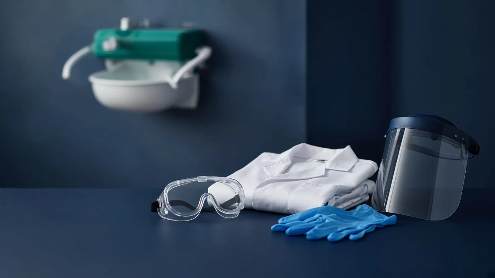
The symptoms show up in two places. Your eyes feel gritty an hour after you cut bands, or the fragment you excised refuses to ligate and the only unusual step was a long look at the gel with the shield up. Both can come from the same transilluminator. Ultraviolet light damages skin and eyes. It also damages the DNA you meant to clone. The shield and the interlock are the controls. A troubleshooting session starts from the symptom, then checks whether those controls were actually in the path of the light.
The gel that put you in front of the lamp is explained in agarose gel electrophoresis for DNA. The stain that usually requires the lamp is in ethidium bromide and safer dye choices still need care. The bench context is biosafety basics for research benches. Imaging accessories and protective eyewear are classes in laboratory consumables. A laboratory specifying an imager can use academic research and a quotation request.
Symptoms first
Gritty eyes, tearing, or pain in bright light, starting later rather than during the session, are the classic pattern people report after an ultraviolet exposure to the eye. The next check is mechanical, not heroic. Was the instrument shield down? Did you look in from the side? Was the lamp still on while you reached under the shield to move the gel? Did a plastic "UV-blocking" visor that somebody left on the bench actually belong to this wavelength? Report the exposure the way the institution says. See occupational health if the plan says to. This article will not tell you what drops to use or how long the pain should last.
Redness on the hands, the wrists, or the face after repeated gel cutting is a skin symptom of the same lamp. The next check is whether any skin was uncovered beside the shield, and whether the assessment required gloves and a coat that actually cover the wrists. A cotton lab coat is not an ultraviolet shield unless the assessment has accepted a specific material for that purpose. Do not diagnose a burn from a paragraph. Do record that the skin was exposed, and do stop using the open lamp.
A band that looks right on the photograph and then will not clone is a DNA symptom. Ultraviolet light, especially the shorter wavelengths used on some transilluminators, creates damage that ligation and transformation will reveal later. The next check is how long the fragment sat on the lamp, whether you used a cutting setting or left the analytical lamp on, and whether the shield was up so you could see better. Then check the cloning controls that have nothing to do with light: the vector backbone, the ligation conditions, and the competent cells. The lamp is a likely cause when the exposure was long. It is not an excuse to skip the other checkpoints in plasmid cloning from insert to colony.
No bands, with a lamp that looks dim or the wrong colour, is an instrument symptom. The stain and the wavelength have to match. An ethidium gel on a blue box, or a blue-light dye on an ultraviolet box with the wrong filter, produces a blank that people try to fix by removing shields. The next check is the dye's excitation, written on its sheet, against the lamp that is actually installed. A dim lamp can also be age. Replacing a lamp is a maintenance task. Bypassing the shield while you wait for a replacement is not the maintenance.
A shield that does not sit down, a cracked shield, or a lamp that stays on when the shield is raised means the control is broken. The next check is to switch the instrument off, label it out of service, and tell the person who owns the equipment. Do not tape a switch. Do not prop the shield. This page will not describe how interlocks are defeated, because that description would be a method for deleting the control.
What the lamp is doing
A transilluminator shines upward through the gel. Common laboratory lamps sit in the ultraviolet-B region, often near 302 nanometres, or in the ultraviolet-C region near 254 nanometres. The shorter wavelength is harsher to DNA and to tissue. Both are ultraviolet. Both are the reason a shield exists. These figures identify lamp classes. They are not a safe dose, and they are not permission to calculate an exposure time from a blog.
The light that reaches the eye can inflame the cornea. The light that reaches the skin can burn it. The light that reaches the DNA creates lesions, including damage that later shows up as a failed clone or as mutations you did not plan. You do not have to choose which harm to believe. The same photons do all three, in different targets. Less time on the lamp reduces all three. The shield blocks the path to you while you look. It does not, by itself, protect the DNA in the gel. DNA protection is time, intensity, and wavelength, or a different imaging system.
The interlock, where the instrument has one, cuts the lamp when the shield opens. It is the control that still works when somebody forgets. If it has been defeated, the instrument is not the instrument the risk assessment described. Put that in the assessment's language: a modified safety device is a change of equipment, and the work stops until a competent person restores it. Risk assessment is a local document is the standard for that sentence.
Biological biosafety documents sit beside this hazard, not instead of it. The WHO laboratory biosafety manual and the CDC BMBL govern the biological material in the lane. The ultraviolet lamp is a physical hazard, and the stain is a chemical hazard under the safety data sheet and, where it applies, the OSHA laboratory standard. A non-pathogenic plasmid does not make the lamp mild.
A session that keeps the controls in place
Turn the lamp on only when the shield is down and you are ready to look. Find the bands, photograph if that is the record, and turn the lamp off. If you must cut, work for the shortest time the fragment allows, with the shield between your face and the gel, and with the blade discipline in sharps and glass Pasteur pipettes. Do not hover. If the band is faint, fix the gel rather than the shield. Percentage, load, and stain are the electrophoresis article's levers.
The branch when you need a picture and a clone from the same lane is to separate the two jobs. Photograph under the controls. For the preparative lane, minimise ultraviolet time, or use a system the assessment has accepted that does not use ultraviolet light. Do not do the photography with the shield up "so the camera sees it" unless the instrument is built for that camera and the assessment says the operator is protected. A phone camera held over an open lamp is a face in the beam.
Blue-light imagers, paired with a dye that those imagers can excite, are the branch when the laboratory wants to stop using ultraviolet light for routine gels. Specify the pair. Keep the chemical waste rule for the dye. Treat a very bright blue source according to the instrument manual. Do not call the box harmless because the light looks familiar.
| Symptom | Likely cause | Next check |
|---|---|---|
| Delayed gritty or painful eyes | Ultraviolet reached the eye around or under a shield | Shield position, side glances, interlock. Report the exposure. No self-experiment |
| Skin redness on hands or face | Uncovered skin near the lamp | What the assessment required for cover. Stop using an open lamp |
| Right-sized band, failed clone | DNA damaged by a long ultraviolet exposure | Time on the lamp, wavelength, whether a blue-light option exists. Check other cloning controls too |
| No bands, dim or wrong-coloured lamp | Wavelength does not match the stain, or the lamp is old | Dye sheet versus installed lamp. Do not remove the shield to compensate |
| Lamp on with the shield open | Interlock failed or was bypassed | Instrument out of service. Do not improvise a workaround |
| Cracked or loose shield | The control is damaged | Stop. A borrowed intact shield from another model may not fit this lamp |
Failure modes that look like skill
People learn to "just glance" with the shield up because the orange plastic makes the band harder to see at an angle. The glance is the exposure. If the shield's plastic is too scratched or too dark to see through, the shield is due for replacement. A scratched shield that you work around is a failed control, the same way fogged goggles are a failed control in eye protection and a face shield.
A second failure is shared instruments. The transilluminator in the gel room serves every group, so nobody owns the cracked hinge. The receiving user still owns the decision to switch it on. If the shield does not close, you do not get a turn. Report it. Cutting the band in the open because the seminar starts in ten minutes is how a scheduling problem becomes an eye injury and a dead clone.
A third failure is spill and lamp together. A gel breaks on the glass, stain side down. You now have a chemical spill of whatever dye you used, on a hot ultraviolet surface. Turn the lamp off before you address the spill, so you are not leaning into the beam, and then follow the stain's spill plan. The identity of the dye still governs the cleanup. The lamp does not sterilise the stain, and it is not a reason to wipe blindly. Spill families are separated in spill cleanup starts with the substance identity.
What this does not decide
The page does not treat your eyes, approve a biosafety level, or clear a modified instrument. Authorisation for the biological work and for the chemical stain sits with the institution. If your eyes hurt, follow the emergency plan. Do not use a troubleshooting table as ophthalmology. Risk assessment and any return-to-work decision sit with the institution, not with a successful ligation the next day.
A borrowed box
Power cuts matter less here than borrowed equipment. A transilluminator from another department can have a different wavelength, a missing interlock, and a shield that does not match the hinge. Heat and humidity cloud and craze plastics, so a shield that was clear last monsoon may now be a reason people lift it. Look at the shield before you look at the gel. Specify, in the room's assessment, which instrument is cleared, and keep a broken one labelled. On a quotation request, ask for the wavelength, whether a shield and an interlock are part of the instrument, and whether a blue-light model exists for the dye you have already been allowed to use. Ask for replacement shields as a named part. Do not ask a seller to confirm that a visor makes an open lamp acceptable.
Questions from the bench
Why do my eyes hurt hours after I looked at a gel?
Ultraviolet light from a transilluminator can inflame the cornea. The pain is often delayed, so the session felt fine and the symptoms arrive later. That pattern is a reason to report the exposure as your institution directs and to have the instrument checked. This page is not a diagnosis and not a treatment. Do not go back to the lamp the same day to see if it happens again.
Does a face shield replace the transilluminator's own shield?
No. The instrument shield and its interlock are the controls built for that lamp. Personal eyewear can be what the assessment adds, and it does not licence you to work with the instrument shield up. If the lamp stays on while the shield is open, the interlock has failed. Take the instrument out of service and report it.
The band was the right size and the cloning failed. Could the lamp be why?
Yes, it could. Ultraviolet light damages DNA, and a long look at a fragment you then excise can nick it enough to ruin a ligation even when the band position was correct. Shorten the exposure, use the intensity the instrument provides for cutting rather than for photography if those settings differ, or move to a blue-light system if the stain and the assessment allow it. Confirm the rest of the cloning checkpoints as well. The lamp is one cause, not the only one.
Is a blue-light transilluminator free of hazard?
It removes the ultraviolet wavelengths when it truly emits only visible blue light, which is a real improvement for eyes, skin, and DNA. The light can still be very bright, and the stain in the gel is still a chemical. Read the instrument instructions and the dye's safety data sheet. Safer illumination is not a reason to skip the chemical waste rule.
References
Manufacturer names identify published method classes. Trademarks remain with their owners. Catalogue records on this site are independent references for enquiry. They are not a statement of inventory, distribution rights or a supply commitment. This page is educational. It is not medical advice, a diagnostic protocol or a biosafety approval.
Catalogue
Related products and categories
These links follow the subject of the article into published manufacturer references. A listing is a reference for an enquiry, not a statement of stock or distribution rights.
Continue in this cluster
Related reading
Biosafety basics for research benchesResearch-bench biosafety is a stack of barriers chosen by a risk assessment. This page explains the stack. It does not approve the work. Agarose gel electrophoresis for DNAHow agarose percentage, buffer and a size ladder turn a DNA sample into a band, and what that band does and does not prove about identity.A glossary of biosafety termsPairs that share a bench still do different jobs. Use this comparison to choose the right word for a cabinet, a chemical, a level, or a control.